Volume 1
The science and practice of medicine / By William Aitken ... From the 4th London ed., with additions, by Meredith Clymer.
- William Aitken
- Date:
- 1866
Licence: Public Domain Mark
Credit: The science and practice of medicine / By William Aitken ... From the 4th London ed., with additions, by Meredith Clymer. Source: Wellcome Collection.
Provider: This material has been provided by the University of Massachusetts Medical School, Lamar Soutter Library, through the Medical Heritage Library. The original may be consulted at the Lamar Soutter Library at the University of Massachusetts Medical School.
937/972 page 927
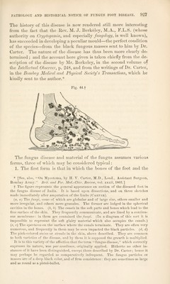
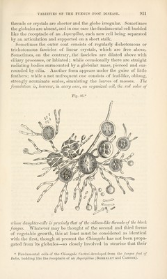
![The history of this disease is now rendered still more interesting from the feci that the Rev. M. J. Berkeley, M.A., F.L.S. (whose authority on Qryptogamia, and especially fungology, is well known), has succeeded in developing a peculiar mould—the perfect condition of the species—from the black fungous masses sent to him by Dr. Carter. The nature of the disease has thus been more clearly de- termined ; and the account here given is taken chiefly from the de- scription of the disease by Mr. Berkeley, in the second volume of the Intellectual Observer, p. 248, and from the writings of Dr. Carter, in the Bombay Medical and Physical Society's Transactions, which he kindly sent to the author.* Fig. 44.f The fungus disease and material of the fungus assumes various forms, three of which may be considered typical: 1. The first form is that in which the bones of the foot and the * [See, also, On Mycetoma, by H. Y. Carter, M.D., Lond., Assistant Surgeon, Bombay Army. Brit, and For. Med.-Chir. Review, vol. xxxii, 1863.] f The figure represents the general appearance on section of the diseased foot in tbe fungus disease of India. It is based upon dissections, and on three sketches made immediately after amputation of the limbs (Carter). (a, a) The fungi, some of which are globular and of large size, others smaller and more irregular, and others mere granules. The former are lodged in the spherical cavities in the bones. (b, b) The canals in the soft parts and bones which lead to the free surface of the skin. They frequently communicate, and are lined by a continu- ous membrane: in them are contained the fungi. (In a diagram of this sort it is impossible to represent the soft glairy material which also occupies the canals.) (c, c) The apertures on the surface where the canals terminate. They are often very numerous, and frequently in them may be seen impacted the black particles, (d, d) The pink-colored stains or streaks in the skin, above described. They are common to both varieties of the disease, and by them it is supposed the growth is multiplied. It is to this variety of the affection that the term fungus disease, which correctly expresses its nature, was par excellence, originally applied. Hitherto no other in- stances of it have been distinguished, except those described by Dr. Carter; hence it may perhaps be regarded as comparatively infrequent. The fungus particles or masses are of a deep black color, and of firm consistence : they are sometimes as large and as round as a pistol-bullet.](https://iiif.wellcomecollection.org/image/b21196606_sciencepracticeo00aitk_0937.jp2/full/800%2C/0/default.jpg)