Hog cholera : its history, nature and treatment, as determined by the inquiries and investigations of the Bureau of animal industry.
- Bureau of Animal Industry
- Date:
- 1889
Licence: Public Domain Mark
Credit: Hog cholera : its history, nature and treatment, as determined by the inquiries and investigations of the Bureau of animal industry. Source: Wellcome Collection.
64/242 page 58
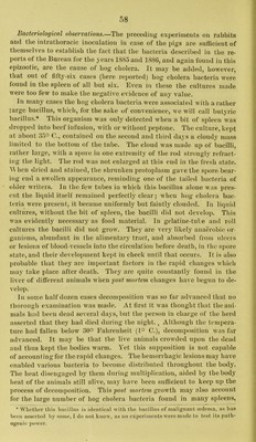
![Bacteriological observations.—T)iq preceding experiments on rabbits and tlie intratlioracic inoculation iu case of the pigs are sufficient of themselves to establish the fact that the bacteria described in the re- ports ot the Bureau for the years 1885 and 1886, and again found in this epizootic, are the cause of hog cholera. It may be added, however, that out of fifty-six cases (here reported) hog cholera bacteria were loiiud iu the spleen of all but six. Even iu these the cultures made were too few to make the negative evidence of any value. In many cases the hog cholera bacteria were associated with a rather large bacillus, which, for the sake of convenience, we will call butyric bacillus.* This organism was only detected when a bit of spleen was dropped into beef infusion, with or without peptone. The culture, kept at about 35° C., contained on the second and third days a cloudy mass limited to the bottom of the tube. The cloud was made up of bacilli, rather large, with a spore in one extremity of the rod strongly refract- ing the light. The rod was not enlarged at this end in the fresh state. A\ hen dried and stained, the shrunken protoplasm gave the spore bear- ing end a swollen appearance, reminding one of the tailed bacteria of • older waiters. In the few tubes in which this bacillus alone was pres- ent the liquid itself remained perfectly clear j when liog cholera bac- teria were present, it became uniformly but faintly clouded. In liquid cultures, without the bit of spleen, the bacilli did not develop. This was evidently necessary as food material. In gelatine-tube and roll cultures the bacilli did not grow. They are very likely’ anaerobic or- ganisms, abundant in the alimentary tract, and absorbed from ulcers or lesions of blood-vessels into the circulation before death, in the spore state, and their development kept in check until that occurs. It is also probable that they arc important factors in the rapid changes which may take place after death. They arc quite constantly found in the liver of different animals when gost mortem changes ha^ e begun to de- velop. In some half dozen cases decomposition was so far advanced that no thorough examination was made. At first it was thought that the ani- mals had been dead several days, but the person in charge of the herd asserted that they had died during the night., Although the tempera- ture had fallen below 30^ Fahrenheit (i° C.), decomposition was far advanced. It may be that the live animals crowded upon the dead and thus kept the bodies warm. Yet this supposition is not capable of accounting for the rapid changes. The hemorrhagic lesions may have enabled various bacteria to become distributed throughout the body. Tlie heat disengaged by them during multiplication, aided by the body lieat of the animals still alive, may have been sufficient to keep up the ])rocess of decomposition. Thispo.vt growth may also account for the large number of hog cholera bacteria found in many spleens, * Whctlier tliis bjicillus is identical ^vitll the bacillus of malignant oideina, as lias been asserted by sonu'., I do not know, as no experiments were made to test its inith- ogenie power.](https://iiif.wellcomecollection.org/image/b28114528_0064.jp2/full/800%2C/0/default.jpg)




 No text description is available for this image
 No text description is available for this image
 No text description is available for this image