An introduction to the study of embryology / by Alfred C. Haddon.
- Alfred Cort Haddon
- Date:
- 1887
Licence: Public Domain Mark
Credit: An introduction to the study of embryology / by Alfred C. Haddon. Source: Wellcome Collection.
Provider: This material has been provided by University of Bristol Library. The original may be consulted at University of Bristol Library.
26/374 page 22
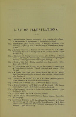
![Fig. 14. Early Stages of Segmkntation of Elysia viridis. Dra^vTi from the living egg. Fig. 15. Segmentation of Oosperm of Frog. [From Balfour after Ecker.] Fig. 16. Gastrulation of Echinus. [A-C original; D and E, from living embryos after Metschnikoff.] Fig. 17, Gastrulation of Fiona nobilis. Fig. 18. Segmentation of Two Prosobranchs : to illustrate the effect of the increase of food-yolk. A.-D. lanthina. [a-g. Fusus after Bobretzki/.] Fig. 19. Syncytial Segmentation of Peripatus. [After Sedgtvick] Fig. 20. Segmentation of Oosperm of Callianassa subterranea. [After Mereschkoioski.] Fig. 21. Blastula and Gastrula of Fresh-water Crayfish (Astacus fluviatilis). [After Reichenhach and Huxley.1 Fig. 22. Figures Illustrating the Development of the Archentebon AND Origin of Mesoblast of Astacus. [From T. J. Parker after Heichenbach.] Fig. 23. Blastula and Gastrula of Amphioxus. [Frotn Claus after Hatschek.] Fig. 24. Blastula and Gastrula Stages of the Frog (Rana temporaria) [After Gotte.] Fig. 25. Three Diagrammatic Longitudinal Sections through an Ideal Type of Vertebrate Embryo, Intermediate in the Mode of Formation of its Layers between Amphibia or Lamprey and Elasmobranchil [From Balfour.] Fig. 26. Longitudinal Section through the Blastoderm of an Elas- mobranch during Gastrulation. {Modifiedfrom Balfour.] Fig. 27. Surface Views of Six Stages in the Segmentation of a Fowl's Oosperm. [From Kdlliker after Coste.] Fig. 28. Semi-Diagrammatic Section through a Fowl's Blastoderm CORRESPONDING TO No. 3, Fig. 27. [Modified from Kdlliker (A) ayid Balfour, B, C] Fig. 29. Section through the Blastoderm of an Unfertilised newly- laid Fowl's Ovum in the Blastula Stage. [After Duval] Fig. 30. Semi-Diagrammatic Section through the Germinal Disc of a Fowl during the Later Stages of Segmentation. Fi<'. ■^i. Portion of a Section through an Unincubated Fowl's Oosperm. [From Klein.] Fig. 32. Section through the Blastoderm of an Unfertilised Canary's Ovum in the Gastrula Stage. [After Duval] Fig. 33. Surface View of the Pellucid Area of the Blastoderm of A Fowl of Twenty Hours. Magnified 24 diameters. [From Kdlliker after His.] Fig. 34. Transverse Section through the J^nterior End of the Primitive Streak of a Fowl's Blastoderm about the Age of Fig. 34. [From Balfour.]](https://iiif.wellcomecollection.org/image/b21443919_0026.jp2/full/800%2C/0/default.jpg)


 No text description is available for this image
 No text description is available for this image
 No text description is available for this image