An introduction to the study of embryology / by Alfred C. Haddon.
- Alfred Cort Haddon
- Date:
- 1887
Licence: Public Domain Mark
Credit: An introduction to the study of embryology / by Alfred C. Haddon. Source: Wellcome Collection.
Provider: This material has been provided by University of Bristol Library. The original may be consulted at University of Bristol Library.
71/374 page 37
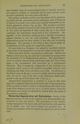
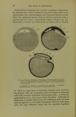
![bating for an hour or two the latter mass is differentiated into a lower°stratum of flattened cells, the hypoblast proper, and scattered mesoblast cells lying between the epiblast and hypoblast. The Fig. 28. — Semi-Diagrammatic Section through a Fowl's Blastoderm corresponding TO No. 3, Fig. 27. [Modi- fled from KoUikcr and Bal- foicr.] B. white yolk spheres; C. iso- lated yellow yolk-sphere. bl. blastoderm; w.y. white yolk, the upper finely granular layer of which is the seat of cell- formation ; y.y. yellow yolk. Fig. A., is cut off just where the white yolk is expanding to form the central mass. imperfect cavity (sub-germinal cavity) between the hypoblast and yolk corresponds with the archenteron of other forms. Duval has figured a longitudinal section of the blastoderm of a Fig. 29.—Section through the Blas- toderm or AN Unfertilised newly- laid Fowl's Ovum in the Blas- tula Stage. lAfter Duval.'] ep. epiblast; n. free nuclei in the yolk, which aggregate protoplasm round themselves to form primitive hypoblast cells; s.c. segmentation-cavity (blasto- ccel). The white yolk is left blank; it rests upon the coarser yellow yolk, re- presented by dots. Canary about this stage (fig. 32). The slit between the blastoderm and the yolk is at the posterior end of the future embryo, and cor- responds with the slit-like archenteric invagination of the Lamprey Fio. 30.—Semi-Diagrammatic Section through the Germinal Disc of a Fowl during THE Later Stages of Segmentation. The central cells are the smallest owing to rapid segmentation ; the large peri- pheral cells of the epiblast rest directly upon the white yolk ; ep. epiblast; 1.1. primitive hypoblast; n. free nuclei in the yolk; w.y. white yolk. (fig. 60) or Frog (fig. 24), or better still, with stage B, or one some- what earlier, of the diagram of the ideal vertebrate (fig, 25). The yolk of the Bird is clearly homologous with the yolk-cells of the Frog; and the surface of the yolk uncovered by the blastoderm](https://iiif.wellcomecollection.org/image/b21443919_0071.jp2/full/800%2C/0/default.jpg)


 No text description is available for this image
 No text description is available for this image
 No text description is available for this image