An introduction to the study of embryology / by Alfred C. Haddon.
- Alfred Cort Haddon
- Date:
- 1887
Licence: Public Domain Mark
Credit: An introduction to the study of embryology / by Alfred C. Haddon. Source: Wellcome Collection.
Provider: This material has been provided by University of Bristol Library. The original may be consulted at University of Bristol Library.
83/374 page 49
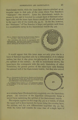
![studied by Merejkowsky, will serve as a type. The segmentation is regular, and results in a large oval blastula, the cells of which are equal in size and ciliated; the wall is also stated to be per- forated by small pores. The embryo next becomes somewhat narrowed at the posterior end.* One by one the cells at the ex- treme hinder end of the embryo become amoeboid and pass into the segmentation-cavity and wander about, congregating at first chiefly at the hinder extremity; eventually the entire segmentation- cavity is filled up by a cellular network formed by the fusion of the pseudopodia of these endoderm.cells. Metschnikoff proposes the name parenchymula for such an embryo, which is formed of an ectodermal layer and a central solid mass of endodermal cells, Fig. 46.—Formation of the Planula of Obelia. '[After Merejkoicski/.]. A. Longitudinal section of a blustula with a few scattered endoderm cells, chiefly at the hind-end. B. Posterior extremity of a slightly earlier sta^e, allow- ing the proliferation of the terminal cell; the resulting endoderm cells immi- grate into the segmontation-caviiy. C. Surface view of a small area of a blastula with two pores. D. Section through a pore. E. Planula in which the segmen- tiition-cavity is filled up with branched endoderm cells. F. Two-layered ciliated planula, with a definite archentric cavity, but no mouth. After a short free life the planula becomes fixed. but without a mouth. The term planula is usually applied to this and the succeeding stage. The endoderm now applies itself to the ectoderm as a definite layer, leaving a central cavity; the archenteron and the free-swimming planula is a ciliated elon- gated two-layered embryo, also destitute of a mouth. After a short free existence, the planula attaches itself by its anterior end, the ectoderm secretes a perisarc, a mouth and tentacles appear, and the hydroid stage commences. In this type the endoderm is formed by immigration, which is positively stated to occur only at one pole of the blastula. W. K. Brooks describes the planula of the Hydromedusoid * The terras anterior and posterior have reference merely to the direction of progression of the larva. D](https://iiif.wellcomecollection.org/image/b21443919_0083.jp2/full/800%2C/0/default.jpg)


 No text description is available for this image
 No text description is available for this image
 No text description is available for this image