An introduction to the study of embryology / by Alfred C. Haddon.
- Alfred Cort Haddon
- Date:
- 1887
Licence: Public Domain Mark
Credit: An introduction to the study of embryology / by Alfred C. Haddon. Source: Wellcome Collection.
Provider: This material has been provided by University of Bristol Library. The original may be consulted at University of Bristol Library.
93/374 page 59
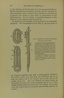
![vary from an apparently epiblastic to an apparently hypoblastic, or to an inter- mediate place of origin. The extreme variations may be neglected, as being in all probability of only secondary significance. Since this was in type Sedgwick has shown that the somites in Peripatus (fig. 69) do not directly arise as archenteric diverticula, but are separated from a pair of mesoblastic bands as in Chaitopoda. The somites are at iirst ventro-Iateral in position, but soon acquire a dorsal extension and divide into two parts. The dorsal parts come into contact above the enteron, but do not unite with their fellows ; anteriorly they are early obliterated, but persist posteriorly as the generative glands. The ventral moieties remain distinct, and consist of a small vesicle situated in the base of the appendages, leading from which is a small coiled tube (hephridium), which acquires an external opening. The Hertwigs have interpreted the formation of the mesoblast in Insects in terms of archenteric diverticula (fig. 55), but the undoubtedly primitive character of Peripatus renders its development especially important. Although the cavities of mesoblastic bands and archenteric diverticula are homologous, their exact relation to one another is somewhat obscure. Whatever vieAvs may be held as to the precise position of the Chsetognatha, Brachiopoda, and Balanoglossus, the presence of archenteric diverticula in these Fig. 56.—Transverse Sections of Embryos of Amphioxus. [A/tei' Hatschel:] A. Section through the first somite or primitive segment of an embryo in which the fifth somite is being formed. B. Section through the same re^^ion of an embi-yo with eight somites. C. Section through the centre of the body of an embryo with eleven somites. al. mesenteron; be. coelom; m. muscle fibres; n. neural plate and canal • nch. notochord. ' forms proves that it occurred in several of the primitive Worms ; so it may be safely concluded that the mesoblast (for the most part, at all events) of the Gephyrea, Polyzoa, and Nematoda belongs to this category. It will probably be shown that mesothelial mesoblast occurs also in all Mollusca. It is probable that the pericardium of this group represents the true body-cavity of other orders; but even if this is the case, there would be a marked preponderance of mesenchyme over mesothelium in the mesoblast. There are not sufficient data to come to a definite conclusion concerning the exact nature of the mesoblast of the Platyhelminths. Origin of the Mesoblast in the Chordata.—There appears to be no valid reason for refusing to accept Bateson's conclusion that Balanoglossus is a persistent representative of an early stage](https://iiif.wellcomecollection.org/image/b21443919_0093.jp2/full/800%2C/0/default.jpg)


 No text description is available for this image
 No text description is available for this image
 No text description is available for this image