On the relations, structure, and function of the valves of the vascular system in Vertebrata / by James Bell Pettigrew.
- Date:
- 1864
Licence: Public Domain Mark
Credit: On the relations, structure, and function of the valves of the vascular system in Vertebrata / by James Bell Pettigrew. Source: Wellcome Collection.
Provider: This material has been provided by the Royal College of Physicians of Edinburgh. The original may be consulted at the Royal College of Physicians of Edinburgh.
4/52 (page 762)
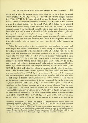
![Structure of the -Veins and Venous Valves. Regarding the composition of the veins, there is, as the reader is aware, some difference of opinion, authorities not being agreed either as to the number or nature of the coats. This may in part be explained by the variation in the thickness of the coats themselves, these, according to John Hunter,* becoming- thinner and thinner in proportion to the size of the vein, the nearer they ap- proach to the heart. In moderate-sized veins an external, a middle, and an internal coat are usually described; the first consisting of cellular, fibrous, and elastic tissue, interlacing in all directions; the second, of waved filaments of areolar tissue, with a certain admixture of non-striped muscular fibres, which run circularly, obliquely, or even longitudinally ; f the third, consisting of one or more strata of very fine elastic tissue, minutely reticulated in a longitudinal direction, the innermost stratum (when several are present) being lined by epithelium. Of these layers, the second and third, from the fact of their contri- buting to the formation of the venous valves, are the most important. The coats of the veins, as has been long known, are tough, elastic, and possessed of con- siderable vital contractility. Of these qualities, the toughness prevents undue dilatation of the vessel when distended with blood; the elasticity and vital con- tractility assisting the onward flow of that fluid, and tending to approximate the segments of the valves, by contracting in the direction of the axis of the vessel. As the valves of the veins are very ample and very flexible, they readily accom- modate themselves to the varying conditions in which they are placed by the elasticity and contractility of the vessel, and by the reflux of the blood. The valves of the veins vary as regards the number of the segments com- posing them, and also slightly as regards structure. In the smallest veins, and where small veins enter larger ones (Plate XXVIII. fig. 0 b), one segment only is present. In middle-sized veins, as they occur in the extremities, two segments (Plate XXVIII. figs. 3, 4, and 7 ab), are usually met with;]; while in the larger veins, as in the internal jugular of the horse, three, and even four segments (Plate XXVIII. figs. 1 and 2, abc,fgh), are by no means uncommon.§ The segments, whatever their number, are semilunar in shape [] (Plate XXVIII. * Hunter on the Blood, pp. 180, 181. I Dr Chevers says, that in the deep as well as in some of the superficial veins of the trunk and neck, the middle coat is composed of several layers of circular fibres, with only here and there a few which take a longitudinal coxirse ; while the middle coat of the superficial and deep veins of the limbs consists of a circular layer, and immediately within this of a strong layer of longitudinal fibres.—Med. Gazette, 1845, p. 638. J In the heart of the frog-fish, sun-fish, sturgeon, American devil-fish, python, and crocodile, a semilunar valve, consisting of two segments, guards the orifice of communication between the sinus venosus and the right auricle. § When four segments are present, two are usually more or less rudimentary (Plate XXVIII. fig. 2/<jr). || John Hunter in speaking of the form of the venous valves, says, their free edges are cut off straight, and are not curved as in the arteries. This, however, is not the case; as may be seen by](https://iiif.wellcomecollection.org/image/b21956200_0006.jp2/full/800%2C/0/default.jpg)