A treatise on the diseases of the eye / by W. Lawrence.
- Sir William Lawrence, 1st Baronet
- Date:
- 1854
Licence: Public Domain Mark
Credit: A treatise on the diseases of the eye / by W. Lawrence. Source: Wellcome Collection.
Provider: This material has been provided by the Francis A. Countway Library of Medicine, through the Medical Heritage Library. The original may be consulted at the Francis A. Countway Library of Medicine, Harvard Medical School.
48/996 (page 38)
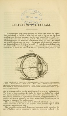
![rays thus refracted ; and certain membranous opaque coverings, surrounding, connecting, and protecting the foregoing parts as well as contributing to their more perfect action. The component textures of the eyeball are usually arranged under the two- fold division of coats and humours. The membranous coverings, and the nerv- ous expansion, constitute the coats, tunics, or membranes : the humours are the transparent media, with the exception of the cornea, which belongs to the coats. The arrangement, although not founded on the clearest grounds, is convenient enough; and we have the further reason for retaining it, of its being established in the language of anatomy by long and universal usage. We must remember, however, that the word humour is used in a merely technical sense, being inap- plicable in its ordinary acceptation of fluid, to the crystalline lens and capsule, and to the vitreous body. The external stratum of the globe is composed of the sclerotica [1, Figs. 1 and 2] and cornea [2, Fig. 1, and d, Fig. 2]; the latter being the anterior transparent part, and the former covering the rest of the eye. When these are removed, we see a membranous covering, distinguished by its deep brown colour, the choroid coat [7, Fig. 1, and 2, Fig. 2], equal in extent to the sclerotica; to this is closely united, in front, the iris, [12, Fig. 1, and tj, Fig. 2], which is placed at a short distance behind the cornea, and perforated near its centre by a round opening called the pupil [13, Fig. 1, and/, Fig. 2], for admitting the light into the interior of the eye; the removal of the choroid exposes the retina, or the nervous expansion. The three coverings just enumerated, i. e., the sclerotica with the cornea, the choroid with the iris, and the retina, are arranged one within the other, concentrically, like the layers of an onion: they may be called respectively the fibrous, vascular, and nervous strata of the eyeball. The humours are three—1st, the vitreous \_m, Fig. 2], which fills the whole concavity of the retina, and forms about four-fifths of the entire bulk of the globe; 2dly, the crystalline, called also crystalline lens [27, Fig. 1, and h, Fig. 2], a nearly spherical body, imbedded in the front of the vitreous humour; 3dly, the aqueous, a small quantity of clear water, filling up the space between the cornea and the front of the crystalline lens, in which space the iris is situated. Only the external or fibrous stratum is complete, that is, closed in all direc- tions ; the vascular layer is perforated in front by the pupil; and the nervous leaves a still greater deficiency, which is filled by the crystalline lens. The membranous layers circumscribe a space, which may be called the cavity of the eyeball, and which is subdivided into three unequal compartments; the posterior, and largest, bounded by the retina, contains the vitreous humour, with the imbedded lens; the two anterior, called the chambers of the eye, are in- closed by the cornea, crystalline, and ciliary processes, separated by the iris, through the pupillary aperture of which they communicate, and filled with the aqueous humour. - Having thus mentioned, in a general way, the parts which make up the globe of the eye, I shall make a few observations on the structure of each, with the view of rendering the description of their morbid affections more intelligible. Sclerotica.—[1, Fig. 1, and 1, Fig. 2.] In dissecting and examining the eyeball, we observe that it does not collapse, but that it retains its figure; this depends on its external investment or sclerotic coat {Albuginea; tunica fibrosa ; Lederhaut, weisse Haul, Germ.). The word sclerotica, which is of Greek extrac- tion (from ax%rtpb$), means hard; the sclerotic coat, therefore, is the firm cover- ing of the eye; it is the most dense, compact, and unyielding texture in the organ. The cornea resembles it in compactness, density, and consequent firm- ness, though it is perfectly transparent, while the sclerotic is entirely opaque. In the older writers the term corneals applied to both these structures; thecornea,](https://iiif.wellcomecollection.org/image/b21063539_0048.jp2/full/800%2C/0/default.jpg)