A treatise on the diseases of the eye / by W. Lawrence.
- Sir William Lawrence, 1st Baronet
- Date:
- 1854
Licence: Public Domain Mark
Credit: A treatise on the diseases of the eye / by W. Lawrence. Source: Wellcome Collection.
Provider: This material has been provided by the Francis A. Countway Library of Medicine, through the Medical Heritage Library. The original may be consulted at the Francis A. Countway Library of Medicine, Harvard Medical School.
52/996 (page 42)
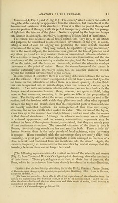
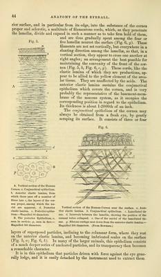
![and mingled with elastic fibrous tissue, flatten out into a membranous form, so as to follow in the main the curvatures of the surfaces of the cornea, and to constitute a series of more than sixty lamellae, intimately united to one another by very numerous processes of similar structure, passing from one to the other, and making it impossible to trace any one lamella, over even a small portion of the cornea. The resulting areolae, which in the sclerotic are irregu- Fig. 3. Vertical section of the Sclerotic and Cornea, showing the continuity of their tissue between the dotted lines, a. Cornea. 6. Sclerotic. In the cornea the tubular spaces are seen cut through, and in the sclerotic the irregular areolas. Cell-nuclei, as at c, are seen scattered throughout, rendered more plain by acetic acid. —Magnified 320 diameters. lar, and on all sides open, are converted in the cornea into tubular spaces, which have a very singular arrangement, hitherto undescribed. They lie in superposed planes, the contiguous ones of the same plane being for the most part parallel, but crossing those of the neighbouring planes at an angle, and sel- dom communicating with them (Fig. 4). The arrangement and size of these Fig. 4. Tubes of the Cornea Proper, as shown in the eye of the Ox by mercurial injection.—Slightly magnified. tubes can be shown by driving mercury, or coloured size, or air, into a small puncture made in the cornea. They may also be shown under a high power by moistening a thin section of a dried cornea, and opening it out by needles. The tissue forming the parietes of these tubes is membranous rather than fibrous, though with the best glasses a fibrous striation may be frequently seen, both in the laminas separating the different series of tubes, and in that dividing those of the same layer from each other. By acetic acid, also, the structure swells, and displays corpuscles resembling those apparent in the white fibrous tissue. Such is the lamellar structure of the cornea, which makes it so much easier to thrust an instrument horizontally than vertically into its substance. The tubes or elongated spaces of which we have spoken, are not distended with any fluid, but are merely moistened in the same way as the areolce of ordinary areolar tissue.]](https://iiif.wellcomecollection.org/image/b21063539_0052.jp2/full/800%2C/0/default.jpg)