A treatise on the diseases of the eye / by W. Lawrence.
- Sir William Lawrence, 1st Baronet
- Date:
- 1854
Licence: Public Domain Mark
Credit: A treatise on the diseases of the eye / by W. Lawrence. Source: Wellcome Collection.
Provider: This material has been provided by the Francis A. Countway Library of Medicine, through the Medical Heritage Library. The original may be consulted at the Francis A. Countway Library of Medicine, Harvard Medical School.
55/996 (page 45)
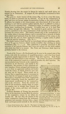
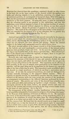
![Vessels shooting into the cornea in disease lie under it, and small ulcers are formed by its destruction. If scraped off, this epithelium is renewed in three or four days. The posterior elastic lamina of the cornea (Fig. 5, d) is a very thin mem- brane, in which no structure can be detected. It has all the transparency of glass, and does not become opaque by maceration, boiling, or the action of acids. It adheres but slightly to the cornea proper, and, when peeled off, it has such a tendency to curl with its anterior surface inwards, that it is difficult to retain a piece of it in an extended form. If floated in water, it exhibits a peculiar glis- tening lustre resulting from its density. It readily tears, yet is so hard that it is bitten through with difficulty. Its elasticity is great, and has been sup- posed to contribute to the exact maintenance of the curvature of the cornea, so necessary for correct vision. This lamina extends only to the circumference of the cornea, where it becomes thinner; and is converted into a network of elastic fibres which bend backwards from the whole circumference of the cornea to the front of the iris, and are there implanted, forming a regular series of pillars around the anterior chambers. (Fig. 13, a.) The elastic lamina is itself lined by an exceedingly delicate epithelium, epithe- lium of the aqueous humour, which exactly resembles that existing on serous membranes (Fig. 5, e, o, p). This epithelium is probably concerned in the secretion of the aqueous humour, but it does not extend over the whole surface with which that fluid is in contact. Drs. Todd and Bowman think that it is probably limited to the cornea.] Vessels of the Cornea.—No bloodvessels can be seen in the normal state of the cornea, even with the assistance of magnifying powers ; and it has been found, in general, equally impracticable to discover them after injection. Both Soem- mering and Eble failed in their attempts. The former has given a beautiful view of the conjunctival vessels in a child six months old, after injection. Nu- merous arteries come forwards on the conjunctiva scleroticae in all directions, and ramify in the membrane: they produce, in fact, numerous mi- nute branches, closely arranged and interwoven, so as to form an apparently uniform red border round the cornea, on which, however, not a sin- gle twig can be traced. These vessels are so minute, that a magnifying power is necessary to prove that the red margin is made up of vessels.1 Eble gives a magnified view of the conjunctival arteries, delineating clearly the superficial and deeper-seated ramifications in the conjunctiva, and on the sclerotica. From the arterial net- work of the membrane, minute twigs, in count- less number, run in straight lines towards the margin of the cornea, and there end abruptly, as if cut off.2 Professor Roemer, of Vienna, has succeeded better in his injections, having filled the arteries Bloodvessels of the Cornea. A. Super- of the conjunctiva corneas in an infant two fi^ai vessels belonging to tho conjunc- months old. He says that the arteries of the tiTal layer> and continued beyond the ocular conjunctiva coming from the lachrymal, m,argin of the cornca' n- VeMnlof the • 111 i j. • jj sclerotic running their course and return- palpebral, and muscular arteries, form a super- ineatthe margin of tho coruca. (From ficial and deeper-seated network on the anterior Toynbee.) Fijr. 7. 1 Iconcs Oculi humani, tab. v. fig. 12. The figure is twice the natural size. 2 Ueber dm Bau und die Krankheiten der Bindchaut des Augapfels, p. 59, tab. ii. fir LI.](https://iiif.wellcomecollection.org/image/b21063539_0055.jp2/full/800%2C/0/default.jpg)