A treatise on the diseases of the eye / by W. Lawrence.
- Sir William Lawrence, 1st Baronet
- Date:
- 1854
Licence: Public Domain Mark
Credit: A treatise on the diseases of the eye / by W. Lawrence. Source: Wellcome Collection.
Provider: This material has been provided by the Francis A. Countway Library of Medicine, through the Medical Heritage Library. The original may be consulted at the Francis A. Countway Library of Medicine, Harvard Medical School.
57/996 (page 47)
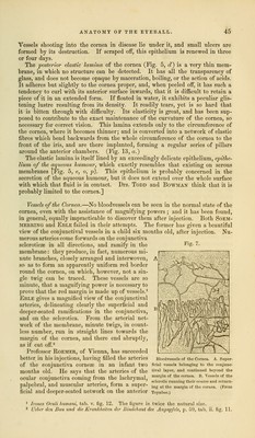
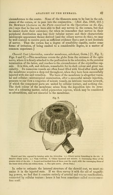
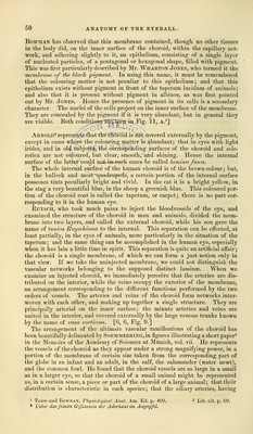
![circumference to the centre. None of the filaments seem to be lost in the sub- stance of the cornea, or to pass into the conjunctiva. {Ibid. Jan. 1846, 231.) Dr. BOWMAN (Lectures on the Parts concerned in the Operations on the Eye, etc.) says that he has not been able to find any nerves in the cornea, but that he cannot doubt their existence; for when we remember that nerves in their peripheral distribution may lose their tubular nature and their characteristic microscopic appearance thence derived (and the ciliary nerves do this), we may be well content to receive pain as sufficient evidence that a part is not destitute of nerves. That the cornea has a degree of sensibility capable, under some forms of irritation, of being exalted to a considerable degree, is a matter of common experience.] Choroid Coat {chorioidea, vascular membrane, aderhaut, Germ.) [7, Fig. 1; Figs. 8 and 9].—This membrane covers the globe from the entrance of the optic nerve, where it is firmly attached to the perforation in the sclerotica, to the anterior termination of the latter, and reaches to the circumference of the crystalline cap- sule. It is thin, soft, and delicate, remarkable for its dark colour and great vas- cularity. Its numerous vessels are often found filled with blood after death, and the membrane acquires a deep red throughout, when they have been successfully injected with size and vermilion. The basis of the membrane is altogether vascu- lar and cellular; microscopical examination, after a successful minute injection, presents to our view a congeries of minute vessels, an intricate vascular network, of which the component parts are united by cellular texture, and nothing else. The dark colour of the membrane arises from the deposition into its struc- ture of a colouring matter, called pigmentum nigrum, which may be considered as adventitious, and not essential to the membrane. Fig. 8. Choroid and Iris, exposed by turning aside the Sclerotica, c, c. Ciliary nerves branching in the iris. d. Smaller ciliary nerve, e, e. Vasa vorticosa. h. Ciliary ligament and muscle, k. Converging fibres of the greater circle of the iris. 1. Looped and knotted form of these near the pupil, with the converging fibros of the lesser circle of the iris within them. o. The optic nerve. (From Zinn.) In order to estimate the internal structure of the choroid coat, we must ex- amine it in the injected state. If we then survey it with the aid of magnify- ing powers, we find that it consists entirely of arterial and venous ramifications, connected by cellular texture: hence it has been sometimes called tunica vascu- tosa oculi.](https://iiif.wellcomecollection.org/image/b21063539_0057.jp2/full/800%2C/0/default.jpg)