A treatise on the diseases of the eye / by W. Lawrence.
- Sir William Lawrence, 1st Baronet
- Date:
- 1854
Licence: Public Domain Mark
Credit: A treatise on the diseases of the eye / by W. Lawrence. Source: Wellcome Collection.
Provider: This material has been provided by the Francis A. Countway Library of Medicine, through the Medical Heritage Library. The original may be consulted at the Francis A. Countway Library of Medicine, Harvard Medical School.
58/996 (page 48)
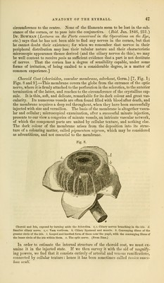
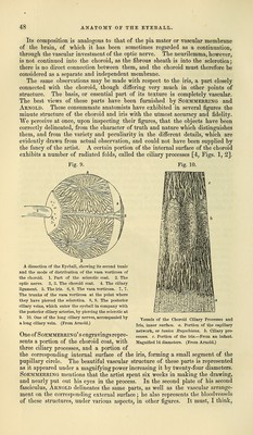
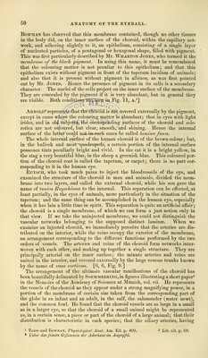
![Its composition is analogous to that of the pia mater or vascular membrane of the brain, of which it has been sometimes regarded as a continuation, through the vascular investment of the optic nerve. The neurilemma, however, is not continued into the choroid, as the fibrous sheath is into the sclerotica; there is no direct connection between them, and the choroid must therefore be considered as a separate and independent membrane. The same observations may be made with respect to the iris, a part closely connected with the choroid, though differing very much in other points of structure. The basis, or essential part of its texture is completely vascular. The best views of these parts have been furnished by Soemmerring and Arnold. These consummate anatomists have exhibited in several figures the minute structure of the choroid and iris with the utmost accuracy and fidelity. We perceive at once, upon inspecting their figures, that the objects have been correctly delineated, from the character of truth and nature which distinguishes them, and from the variety and peculiarity in the different details, which are evidently drawn from actual observation, and could not have been supplied by the fancy of the artist. A certain portion of the internal surface of the choroid exhibits a number of radiated folds, called the ciliary processes [4, Figs. 1, 2]. Fig. 9. Fig. 10. A dissection of the Eyeball, showing its second tunic and the mode of distribution of the vasa vorticosa of the choroid. 1. Part of the sclerotic coat. 2. The optic nerve. 3, 3. The choroid coat. 4. The ciliary- ligament. 5. The iris. 6, 6. The vasa vorticosa. 7, 7. The trunks of the vasa vorticosa at the point where they have pierced the sclerotica. 8, 8. The posterior ciliary veins, which enter the eyeball in company with the posterior ciliary arteries, by piercing the sclerotic at 9. 10. One of the long ciliary nerves, accompanied by a long ciliary vein. (From Arnold.) Vessels of the Choroid Ciliary Processes and Iris, inner surface, a. Portion of the capillary network, or tunica Ruyschiana. b. Ciliary pro- cesses, c. Portion of the iris.—From an infant. Magnified 14 diameters. (From Arnold.) One of Soemmerring' s engravings repre- sents a portion of the choroid coat, with three ciliary processes, and a portion of the corresponding internal surface of the iris, forming a small segment of the pupillary circle. The beautiful vascular structure of these parts is represented as it appeared under a magnifying power increasing it by twenty-four diameters. Soemmerring mentions that the artist spent six weeks in making the drawing, and nearly put out his eyes in the process. In the second plate of his second fasciculus, Arnold delineates the same parts, as well as the vascular arrange- ment on the corresponding external surface; he also represents the bloodvessels of these structures, under various aspects, in other figures. It must, I think,](https://iiif.wellcomecollection.org/image/b21063539_0058.jp2/full/800%2C/0/default.jpg)