A treatise on the diseases of the eye / by W. Lawrence.
- Sir William Lawrence, 1st Baronet
- Date:
- 1854
Licence: Public Domain Mark
Credit: A treatise on the diseases of the eye / by W. Lawrence. Source: Wellcome Collection.
Provider: This material has been provided by the Francis A. Countway Library of Medicine, through the Medical Heritage Library. The original may be consulted at the Francis A. Countway Library of Medicine, Harvard Medical School.
61/996 (page 51)
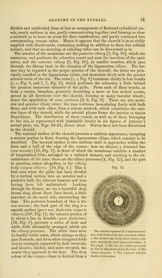
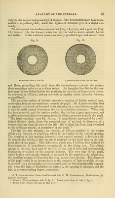
![divided and subdivided, form at last an arrangement of flattened cylindrical ves- sels, nearly uniform in size, partly communicating together, and forming so close a network as to leave no room for finer ramifications, and partly continued into corresponding venous tubes. Hence it appears tbat the choroid is most richly supplied with bloodvessels, containing nothing in addition to these but cellular texture, and that no secreting or exhaling tubes can be discovered in it. The arteries of the membrane are the posterior ciliary [1, Fig. 16], which are numerous, and perforate the sclerotica round and near the insertion of the optic nerve, and the anterior ciliary [3, Fig. 16], in smaller number, which pass through the fibrous tunic in the situation of the ligamentum ciliare. The for- mer may be regarded as the proper arteries of the choroid; the latter are prin- cipally ramified in the ligamentum ciliare, and inosculate freely with the greater arterial circle of the iris. The veins [e, e, Fig. 8] terminate chiefly in four trunks [e, e, Fig. 8, and 7, 7, Fig. 9], which perforate the sclerotica a little behind the greatest transverse diameter of the globe. From each of these trunks, as from a centre, branches, generally describing a more or less arched course, spread in all directions over the choroid, forming so many vascular whorls; hence the appellation of vam vorticosa [6, 6, Fig. 9]. There are also poste- rior and anterior ciliary veins: the vasa vorticosa inosculating freely with both these, and with each other, form a venous network, which constitutes the exte- rior layer of tbe choroid, while the arterial plexus forms the interior or tunica Ruyschiana. The distribution of these vessels, as well as of those belonging to the iris, is represented with inimitable beauty in six figures of Arnold's second plate, viz. Figs. 16-21, Icones Anat. Nerves have not been discovered in the choroid. The external surface of the choroid presents a uniform appearance, excepting a narrow portion in front, forming the ligamentum ciliare, which remains to be described. The internal surface is also uniform until it approaches within two lines and a half of the edge of the cornea: here we observe a dentated line (ora serrata) [11, Fig. 1], in front of which the membrane exhibits a number of longitudinal folds, arranged in a radiated manner, and reaching to the cir- cumference of the lens; these are the ciliary processes [4, Fig. 12], and the part in question, taken altogether, is the ciliary hocly (corpus ciliare). [10, Fig. 1.] This is best seen when the globe has been divided by a vertical section into an anterior and a posterior half, the vitreous humour and lens having been left undisturbed. Looking through the former, we see a beautiful deep brown ring, about three lines broad, a little narrower on the nasal side, surrounding the lens. The posterior boundary of this is the ora serrata; the back part of the ring is a smooth surface (pars nan fimhriata corporis ciliaris) [10', Fig. 1]; the anterior portion of it, about a line in breadth (pars fimhriata) [10, Fig. 1], presents a series of dark and white folds alternately arranged, which are the ciliary processes. The white lines arise by hardly visible striae, which enlarge as they pass forwards, and form white elongated folds closely arranged, separated by dark intervals, and broader, thicker, and more elevated, the nearer they approach to the lens. The deep colour of the corpus ciliare is derived from a Fig. 12. mm v The anterior segment f a transverse sec- tion of the Globe of the Kyc. seen from within. 1. The divided edge of the three tunics; scle- rotic, choroid (thedark layer), and retina. 'J. The pupil. 3. The iris, the surface presented to view in this section being the uvea. t. The ciliary processes. 5, The scalloped anterior border of the retina.](https://iiif.wellcomecollection.org/image/b21063539_0061.jp2/full/800%2C/0/default.jpg)