A treatise on the diseases of the eye / by W. Lawrence.
- Sir William Lawrence, 1st Baronet
- Date:
- 1854
Licence: Public Domain Mark
Credit: A treatise on the diseases of the eye / by W. Lawrence. Source: Wellcome Collection.
Provider: This material has been provided by the Francis A. Countway Library of Medicine, through the Medical Heritage Library. The original may be consulted at the Francis A. Countway Library of Medicine, Harvard Medical School.
65/996 (page 55)
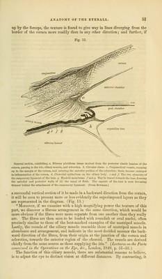
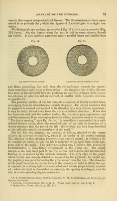
![vary in this respect independently of disease. The Soemmerrings1 have repre- sented it as perfectly flat; while the figures of Arnold2 give it a slight con- vexity. We distinguish two surfaces, an anterior [Fig. 14] (iris), and a posterior [Fig. 15] (uvea). On the former, when the part is laid in water, minute flocculi are visible. It also exhibits numerous nearly parallel larger and smaller lines Pig. 14. '% An anterior view of the Iris. A posterior view of the Iris, or Uvea. and fibres, proceeding like radii from the circumference towards the centre : these sometimes unite so as to form arches. An irregular line divides this sur- face more or less distinctly into two portions; an external, or larger circle (anrm- lus extermis, or ciliaris), and an internal, or smaller circle (an nidus i)iternus, or pupillar is). The posterior surface of the iris presents a number of faintly marked lines, converging from its circumference towards the pupil. Dr. Jacob considers that its pigment is covered and secured in its situation by a very delicate membrane: it may be easily turned down from the iris as a distinct structure. When this has been removed, and the surface washed, the iris has a gray appearance, and exhibits numerous fibres converging from the ciliary processes towards the pupil. The latter opening, says Dr. Jacob, is immediately surrounded by a well- defined distinct circle, about the twentieth part of an inch in diameter, of a denser structure than the rest of the iris; this is what has been long described as the orbicular muscle, or constrictor of the pupil.3 The iris has two margins; an external, or ciliary, attached to the corpus ciliare; an internal, or pupillary, which is the border of the central opening. The situation of this opening, however, is not exactly central; it deviates a lit- tle towards the inside, so that the iris is narrower on the nasal than on the tem- poral side of the pupil. This difference, which was, I believe, first noticed by SOEMMERRING, is immediately recognizable in the living eye. The ciliary margin is the only fixed part of the iris, all the rest is loose and unconnected, floating in or washed by the aqueous humour. The very edge of the pupil, which is thin and sharply defined, is coloured by the pigment; we might say, the pupillary margin is formed by the uvea, rather than the iris. The diameter of the pupil varies in an inverse ratio to the quantity of light to which the eye is exposed : in a strong light it is contracted, and the iris proportionally expand- ed ; when the light is diminished in quantity, the opening is enlarged, and the iris, in a corresponding degree, contracted. ' 8. T. Soemmerrino, Icones Oculi humani, tab. 8. W. Soemmerrino, Dc Oculorum, ,jc Sectione horizontali. 2 Arnold, Untersuchungen, tub. 8, fig. 2. Icones Anal. fasc. ii. tub. 2, fig. 4. 3 Medico- Chir. Trans, vol. xii. p. 513, 541.](https://iiif.wellcomecollection.org/image/b21063539_0065.jp2/full/800%2C/0/default.jpg)