A treatise on the diseases of the eye / by W. Lawrence.
- Sir William Lawrence, 1st Baronet
- Date:
- 1854
Licence: Public Domain Mark
Credit: A treatise on the diseases of the eye / by W. Lawrence. Source: Wellcome Collection.
Provider: This material has been provided by the Francis A. Countway Library of Medicine, through the Medical Heritage Library. The original may be consulted at the Francis A. Countway Library of Medicine, Harvard Medical School.
66/996 (page 56)
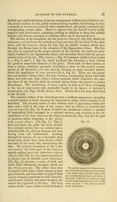
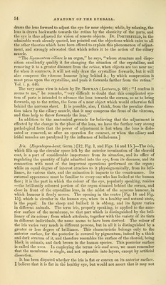
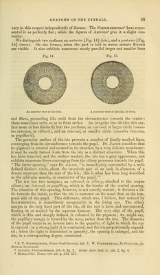
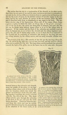
![The notion that the iris is a continuation of the choroid, or, in other words, that these two are parts of one and the same continuous structure, is contradicted, not only by the obvious differences between them in organization and function, but also by the mode of union, by the distinct sources of vascular supply in the two cases, and by the total absence of nerves in the one instance, while the other part is furnished with them as abundantly as any organ in the body. The an- terior circular edge of the ligamentum ciliare, and of the corpus ciliare, forms a groove or plait, in which the outer or ciliary edge, or the greater circumfer- ence of the iris is set; the union not being firm or intimate, but such that the iris can be drawn away without injury either to it or to the ciliary body or ligament. If the cornea and the front of the sclerotica are carefully removed from the human eye, so as to leave the choroid and the ciliary ligament unin- jured, the iris can be drawn away easily, so as to show that it is connected to the choroid, not by continuity of substance, but merely by the long and ante- rior ciliary arteries, by the ciliary nerves, and an easily lacerable cellular text- ure. Bloodvessels of the Iris.—The arteries of the iris are the two long ciliary [2, Fig. 16, and 6, 6, Fig. 20] (the external and internal), and the anterior ciliary, about sis in number [3, Fig. 16]. The long ciliary arteries perforate the sclerotica towards the back of the globe, one on the inner, one on the outer side: they pass Fig. 16. Fig. 17. An enlarged view of the Arteries of the Iris. a. Optic nerve, b. Sclerotic, c. Ciliary ligament, d. Iris. 1. Poste- rior ciliary arteries perforating the sclerotic. 2. Long (exter- nal) ciliary artery. 3. Anterior (short) ciliary arteries. (The figure is larger than natural.) (From Arnold.) forward between the sclerotica and choroid, along the middle of the globe, to the liga- mentum ciliare, where each of them divides into two branches, which run round the greater circumference of the iris, and, unit- ing together, constitute the greater arterial circle [2, Fig. 21] of the iris. The short ciliary arteries pass through the sclerotica in the front of the eye, and join the circle before described. Numerous branches pro- ceed from this circle, and run in a nearly parallel direction along the iris, inosculating in the situation of the lesser circle, so as A segment of the Anterior Face of the Iris with its vessels injected.—Magnified 25 diam- eters. 1,1. A portion of the pupillary circum- ference of the iris. 2, 2. A part of its greater circumference surrounded hy a branch of the long ciliary artery. 3. Part of the lesser circle of the iris. 4, 4. Part of its greater circle. 5, 5. Three arteries which are larger than the others, and coming from the greater circle are lost in the iris. 6. Smaller arteries arising from these. 7. Branches of the larger arteries, which are lost in the smaller circle of the iris. An outline of the natural' size of this piece is seen on the side of the figure.](https://iiif.wellcomecollection.org/image/b21063539_0066.jp2/full/800%2C/0/default.jpg)