A treatise on the diseases of the eye / by W. Lawrence.
- Sir William Lawrence, 1st Baronet
- Date:
- 1854
Licence: Public Domain Mark
Credit: A treatise on the diseases of the eye / by W. Lawrence. Source: Wellcome Collection.
Provider: This material has been provided by the Francis A. Countway Library of Medicine, through the Medical Heritage Library. The original may be consulted at the Francis A. Countway Library of Medicine, Harvard Medical School.
74/996 (page 64)
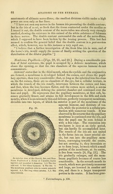
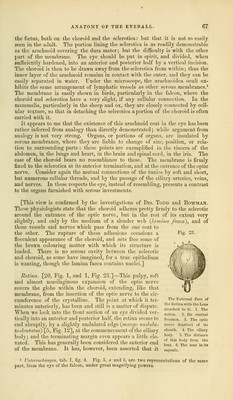
![anastomosis of ultimate nerve-fibres; the smallest divisions visible nnder a high power are seen only as fine lines. I have not seen any nerves in the human iris presenting the double contour; but in the iris of a cat, so fresh that the tissue contracted under the needles as I teased it out, the double contour of the nerve-tubes was already very strongly marked, showing the existence in this animal of the white substance of Schwann in these nerves. The doubie contour surrounded the ends of the nerve-fibres, which I supposed to have been broken by the teasing process. This last fact seemed to confirm the general belief that the double contour is a post-mortem effect, which, however, was in this instance a very rapid one. I believe that a further investigation of the fresh blue iris in man, and of the horse's iris, would supply the means of finally settling the question of the distribution of the dilator pupillse.] Membrana Papillaris.—[Figs. 20, 21, and 22.] During a considerable por- tion of fetal existence, the pupil is occupied by a delicate membrane, which closes the opening, so that the two chambers of the aqueous humour do not communicate. Arnold1 states that in the third month, when the eyelids and the conjunctiva are formed, a membrane is developed behind the cornea, and closes the pupil- lary aperture, then very considerable: that, so long as the spherical lens lies close on the flat cornea, there are no chambers of the eye, but a cellular texture, in which the vessels of the iris ramify, intervenes between the cornea and lens; and that, when the lens becomes flatter, and the cornea more arched, a serous membrane is developed, defining the anterior chamber and continued over the lens and pupil. He represents that the pupillary membrane, at first soft, be- comes gradually firmer, and attains its full development in the fifth and sixth months, when it is an extremely thin and delicate, perfectly transparent membrane, divisible into two layers, of which the anterior is part of the membrane of the aqueous humour, and destitute of ves- sels, while the posterior is a cellular pro- duction with numerous vessels. It was observed by Rtjdolphi that the pupillary membrane is continued over the iris, and that the pupil can be seen behind it with a free edge. The separation into two layers is easy at an early period, but can hardly be accomplished later. The vessels of the iris are not united in the fetus into an inner arterial cir- cle, but run into the pupillary mem- brane, converging towards the centre, but not crossing it; they inosculate so as to form loops, between which a small portion of the membrane, at its centre, is free from vessels [6, Fig. 21]. The iris gradually enlarges, and the mem- brana pupillaris becomes of course less considerable. In the seventh month its vessels, which were previously numerous and large, are lessened in number and size, and there is a larger transparent portion in the centre. It has been gene- An anterior view of the Choroid, Iris, and Mem- hrana Pupillaris of a Fcetus of 7 months, highly injected, and magnified 4 diameters. 1,2. The cho- roid coat. 3. The ciliary ligament. 4. The iris. 5. The memhrana pupillaris with its vessels minutely injected. 6, 6. The long ciliary arteries. 7, 7. The vasa vorticosa. Untersuclmngen, p. 156.](https://iiif.wellcomecollection.org/image/b21063539_0074.jp2/full/800%2C/0/default.jpg)