A treatise on the diseases of the eye / by W. Lawrence.
- Sir William Lawrence, 1st Baronet
- Date:
- 1854
Licence: Public Domain Mark
Credit: A treatise on the diseases of the eye / by W. Lawrence. Source: Wellcome Collection.
Provider: This material has been provided by the Francis A. Countway Library of Medicine, through the Medical Heritage Library. The original may be consulted at the Francis A. Countway Library of Medicine, Harvard Medical School.
76/996 (page 66)
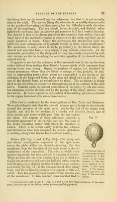
![membrane still preserves its integrity, though perfectly transparent. The period now approaches when it is to disappear; this occurrence takes place, according to my observations, a short time previous or subsequent to birth. In every instance where I have made the examination, I have found the mem- brana pupillaris existing in a greater or less degree of perfection in the new- born infant; frequently perfect, without the smallest breach, sometimes present- ing ragged apertures in_several places, and in other instances, nothing existing but a remnant hanging across the pupil like a cobweb. I have even succeeded in injecting a single vessel in the membrana pupillaris of the ninth month. When I have examined it in subjects who have lived for a week or fortnight after birth, as proved by the umbilicus being healed, I have uniformly found a few shreds still remaining. It is obvious, from the preceding observations, that the membrane does not disappear by a rent taking place in the centre, and re- traction of the vessels to thejiris, as supposed by Blumenbach; but that it at first loses its vascularity, then becomes exceedingly thin and delicate, and is finally absorbed. Portal1 says, that it disappears at the time of birth, or a little later. Arnold2 states, that he had frequently seen some vascular ramifi- cations in the pupil, remains of the pupillary membrane in the eyes of newly- born infants; and that he once saw, in both eyes of a child at full time, the membrane perfect, excepting a small point in the centre, and well supplied with bloodvessels. Arachnoid Coat of the Eye [Membrana Fusca.—5, Fig. 1].—Some anato- mists have recently described a serous membrane, lining the cornea, spread over the choroid, and bearing the same relation to those tunics that the arach- noid does to the dura and pia mater: hence they have called it arachnoidea oculi. It has been observed that there is a fluid between the sclerotica and choroid, giving their surfaces a shining, if not very moist appearance; that minute injections escape here, as they do into serous cavities, and that morbid collections of fluid have been found in this situation. Zinn3 expressly compares the space between the two membranes to the serous cavities; and many anatomists have regarded the external surface of the choroid, or the lining of the sclerotica as serous. Schreiber says sclerotica in facie sua interna naturam membrane serosae pras se fert, sicut chorioidea in facie sua externa.4 Fraenzel5 and Meckel6 consider the internal surface of the sclerotica to be serous, and analogous to the internal surface of the dura mater, while the latter represents this serous layer of the sclerotica as derived from the arachnoid coat of the brain. Although the arachnoid of the eye has escaped the notice of Zinn, of Soem- merring, and of Jacob,7 Arnold8 describes it regularly, and introduces it in his view of the vertically divided globe, both in his Untersuchungen (tab. 3, Fig. 2), and in the Tabulae Anatomicse (tab. 2, Fig. 4). He says that the arachnoidea oculi can be easily exhibited, by careful dissection, in the eye of 1 Memoires du Museum, torn. iv. 2 Lib. cit. p. 158.—The bloodvessels of the membrana pupillaris have been repeatedly delineated: by Blumenbach, Institutiones Physiological, tab. 2. Soemmerring, Icones Oculi Hurnani, tab. 5. J. Cloquet, Memoire sur la Membrane Pupillaire, et sur la formation du petit cercle arteriel de VIris, Paris, 1818. The best representations of the subject are those of Dr. Jacob, Medico-Chirurgical Transactions, vol.xii.pl. 10, and of Arnold, Icon. Anat. fasc. 2, tab. 2, fig. 19. 3 Descrip. Anat. Oculi Humani, p. 25. 4 E,adius, Script. Ophthal. minor, vol. iii. p. 128. See also YYardrop, Morbid Anatomy of the Human Eye, vol. ii. ch. 27 and 53. 5 Ammon's Zeitschrift, vol. i. p. 13. 6 Handbuch der Mensch. Anat. b. iv. p. 73. 7 Article Eye in the Cyclopaedia of Anatomy and Physiology. 8 Untersuchungen, p. 33, et seq.](https://iiif.wellcomecollection.org/image/b21063539_0076.jp2/full/800%2C/0/default.jpg)