A treatise on the diseases of the eye / by W. Lawrence.
- Sir William Lawrence, 1st Baronet
- Date:
- 1854
Licence: Public Domain Mark
Credit: A treatise on the diseases of the eye / by W. Lawrence. Source: Wellcome Collection.
Provider: This material has been provided by the Francis A. Countway Library of Medicine, through the Medical Heritage Library. The original may be consulted at the Francis A. Countway Library of Medicine, Harvard Medical School.
77/996 (page 67)
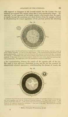
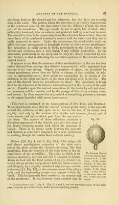
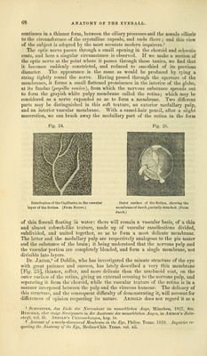
![the foetus, both on the choroid and the sclerotica: but that it is not so easily seen in the adult. The portion lining the sclerotica is as readily demonstrable as the arachnoid covering the dura mater; but the difficulty is with the other part of the membrane. The eye should be put in spirit, and divided, when sufficiently hardened, into an anterior and posterior half by a vertical incision. The choroid is then to be drawn away from the sclerotica from within; thus the inner layer of the arachnoid remains in contact with the outer, and they can be easily separated in water. Under the microscope, the arachnoidea oculi ex- hibits the same arrangement of lymphatic vessels as other serous membranes.1 The membrane is easily shown in birds, particularly in the falcon, whei'e the choroid and sclerotica have a very slight, if any cellular connection. In the mammalia, particularly in the sheep and ox, they are closely connected by cell- ular texture, so that in detaching the sclerotica a portion of the choroid is often carried with it. It appears to me that the existence of this arachnoid coat in the eye has been rather inferred from analogy than directly demonstrated; while argument from analogy is not very strong. Organs, or portions of organs, are insulated by serous membranes, where they are liable to change of size, position, or rela- tion to surrounding parts : these points are exemplified in the viscera of the abdomen, in the lungs and heart, in the brain and spinal cord, in the iris. The case of the choroid bears no resemblance to these. The membrane is firmly fixed to the sclerotica at its anterior termination, and at the entrance of the optic nerve. Consider again the mutual connections of the tunics by soft and short, but numerous cellular threads, and by the passage of the ciliary arteries, veins, and nerves. In these respects the eye, instead of resembling, presents a contrast to the organs furnished with serous investments. [This view is confirmed by the investigations of Drs. Todd and Bowman. These physiologists state that the choroid adheres pretty firmly to the sclerotic around the entrance of the optic nerve, but in the rest of its extent very slightly, and only by the medium of a slender web {lamina fused), and of those vessels and nerves which pass from the one coat to the other. The rupture of these adhesions occasions a Fig. 23. flocculent appearance of the choroid, and sets free some of the brown colouring matter with which its structure is loaded. There is no serous cavity between the sclerotic and choroid, as some have imagined, for a true epithelium is wanting, though the lamina fusca contains nuclei.] Retina. [20, Fig. 1, and 1, Fig. 23.]—This pulpy, soft and almost mucilaginous expansion of the optic nerve covers the globe within the choroid, extending, like that membrane, from the insertion of the optic nerve to the cir- cumference of the crystalline. The point at which it ter- minates anteriorly, has been and still is a matter of dispute. When we look into the front section of an eye divided ver- tically into an anterior and posterior half, the retina seems to end abruptly, by a slightly undulated edge {margo undula- to-dentatus) [5, Fig. 12], at the commencement of the ciliary body; and the terminating margin even appears a little ele- vated. This has generally been considered the anterior end of the membrane. It has, however, been asserted that it The External Face of the Retina with the Lens attached to it. 1. The retina. 2. Its central foramen. 3. Tho optic nerve deprived of its sheath. 4. Tho ciliary body. 5. Tho distanco of this body from the lens. 6. Tho lens in its capsulo. 1 Untersuchungcn, tab. 1, fig. 4. Fig. 5, a and b, are two representations of the same part, from the eye of the falcon, under great magnifying powers.](https://iiif.wellcomecollection.org/image/b21063539_0077.jp2/full/800%2C/0/default.jpg)