A treatise on the diseases of the eye / by W. Lawrence.
- Sir William Lawrence, 1st Baronet
- Date:
- 1854
Licence: Public Domain Mark
Credit: A treatise on the diseases of the eye / by W. Lawrence. Source: Wellcome Collection.
Provider: This material has been provided by the Francis A. Countway Library of Medicine, through the Medical Heritage Library. The original may be consulted at the Francis A. Countway Library of Medicine, Harvard Medical School.
78/996 (page 68)
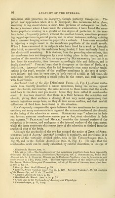
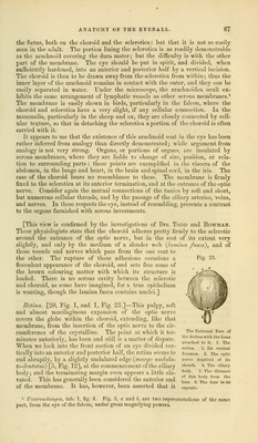
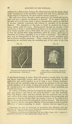
![continues in a thinner form, between the ciliary processes and the zonula ciliaris to the circumference of the crystalline capsule, and ends there; and this view of the subject is adopted by the most accurate modern inquirers.1 The optic nerve passes through a small opening in the choroid and sclerotic coats, and here a singular circumstance is observed. If we make a section of the optic nerve at the point where it passes through these tunics, we find that it becomes suddenly constricted, and reduced to one-third of its previous diameter. The appearance is the same as would be produced by tying a string tightly round the nerve. Having passed through the aperture of the membranes, it forms a small flattened prominence in the interior of the globe, at its fundus {papilla conica), from which the nervous substance spreads out to form the grayish white pulpy membrane called the retina; which may be considered as a nerve expanded so as to form a membrane. Two different parts may be distinguished in this soft texture, an exterior medullary pulp, and an interior vascular membrane. With a camel-hair pencil, after a slight maceration, we can brush away the medullary part of the retina in the form Fig. 24. Fig. 25. Distribution of the Capillaries in the vascular layer of the Retina. (From Berres.) Outer surface of the Retina, showing the membrane of Jacob, partially detached. (From Jacob.) of thin flocculi floating in water: there will remain a vascular basis, of a thin and almost cobweb-like texture, made up of vascular ramifications divided, subdivided, and united together, so as to form a most delicate membrane. The latter and the medullary pulp are respectively analogous to the pia mater and the substance of the brain; it being understood that the nervous pulp and the vascular portion are completely blended, and form a single membrane, not divisible into layers. Dr. Jacob,3 of Dublin, who has investigated the minute structure of the eye with great patience and success, has lately described a very thin membrane [Fig. 25], thinner, softer, and more delicate than the arachnoid coat, on the outer surface of the retina, giving an external covering to the nervous pulp, and separating it from the choroid, while the vascular texture of the retina is in a manner interposed between the pulp and the vitreous humour. The delicacy of this structure, and the consequent difficulty of demonstrating it, will account for differences of opinion respecting its nature. Arnold does not regard it as a 1 Schneider, das Ende der Nervenhaut im mensehlichen Auge, Munch en, 1827, 4to. Huschke, uber einige Streitpuncte in der Anatomie des mensehlichen Auges, in Ammon's Zeits- chrift, vol. iii. Arnold's Untersuchungen, kap. iv. 2 Account of a newly-discovered Membrane in the Eye, Philos. Trans. 1819. Inquiries re- specting the Anatomy of the Eye, Medico-Chir. Trans, vol. xii.](https://iiif.wellcomecollection.org/image/b21063539_0078.jp2/full/800%2C/0/default.jpg)