Remarks on the anatomy of the knee-joint / by Holmes Coote.
- Coote, Holmes, 1817-1872.
- Date:
- [1850]
Licence: Public Domain Mark
Credit: Remarks on the anatomy of the knee-joint / by Holmes Coote. Source: Wellcome Collection.
Provider: This material has been provided by The Royal College of Surgeons of England. The original may be consulted at The Royal College of Surgeons of England.
2/6 page 76
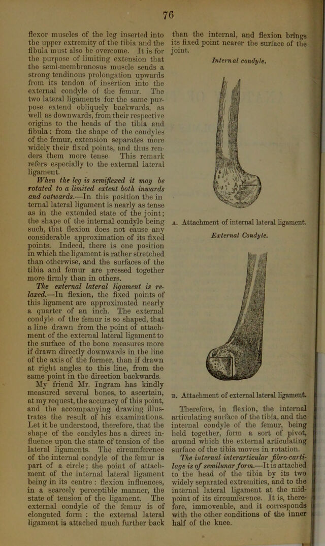
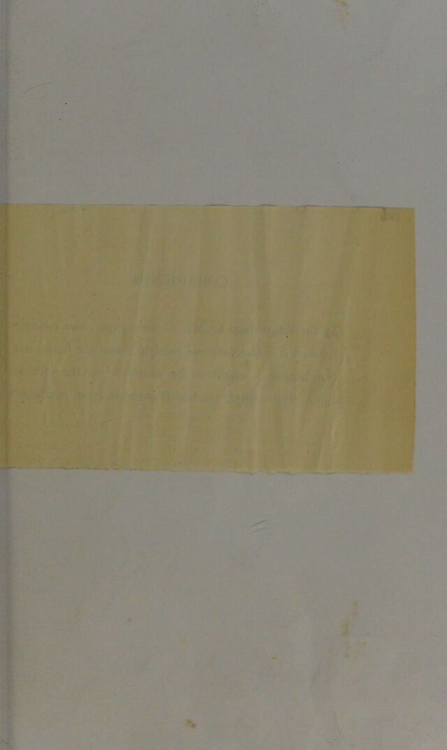
![76 flexor muscles of the leg inserted into the upper extremity of the tibia and the fibula must also be overcome. It is for the purpose of limiting extension tliat the semi-memhranosus muscle sends a strong tendinous prolongation upwards from its tendon of insertion into the external condyle of the femur. Tlie two lateral ligaments for the same pur- pose extend obliquely hachwards, as well as downwai’ds, from their res])ecti ve origins to the heads of the tibia and fibula: from the shape of the condyles of the femur, extension separates more widely their fi.xed points, and thus ren- ders them more tense. This remark refers especially to the external lateral ligament. When the leg is semijlexed it may be rotated to a limited extent both inwards and outwards.—In this position the in temal lateral ligament is nearly as tense as in tlie extended state of the joint; the shape of the internal condyle being such, that flexion does not cause any considerable approximation of its fixed points. Indeed, there is one position in which the ligament is rather stretched than otherwise, and the surfaces of the tibia and femm- are pressed together more firmly than in others. The external lateral ligament is re- laxed.—In flexion, the fixed points of this ligament are approximated nearly a quarter of an inch. The external condyle of the femur is so shaped, that a line drawn from the point of attach- ment of the external lateral ligament to the siuface of the bone measures more if drawn dfrectly downwards in the line of the axis of the former, tlian if di-awn at right angles to this line, fr’om the same point in the dh-ection backwards. My friend Mr. Ingram has kindly measured several bones, to ascertain, at my request, the accmacy of this point, and the accompanying drawing illus- trates the result of his examinations. Let it be understood, therefore, that the shape of the condyles has a direct in- fluence upon the state of tension of the lateral ligaments. The circumference of the internal condyle of the femm' is part of a circle; the point of attach- ment of the internal lateral ligament being in its centre : flexion influences, in a scarcely perceptible manner, the state of tension of the ligament. The external condyle of the femm' is of elongated foi-m : the external lateral ligament is attached much fm-thor back than the internal, and flexion brings its fixed point nearer tlie sm-lace of the joint. Internal condyle. A. Attachment of internal lateral ligament. External Condyle. B. Attachment of external lateral ligament. Tlierefore, in flexion, the internal articulating surface of the tibia, and the internal condyle of the femur, being held together, form a sort of pivot, around which the external articulating surface of the tibia moves in rotation. The internal interartieular jibro-carti- luge is of semilunar form.—It is attached to the head of the tibia by its two widely separated exti'emities, and to the internal lateral bgament at the mid- point of its circumference. It is, there- fore, immoveable, and it con'esponds with the other conditions of the inner half of the knee. I](https://iiif.wellcomecollection.org/image/b22424611_0004.jp2/full/800%2C/0/default.jpg)