Text-book of human physiology : including histology and microscopical anatomy with especial reference to the practice of medicine / by L. Landois.
- Leonard Landois
- Date:
- [1904], ©1904
Licence: Public Domain Mark
Credit: Text-book of human physiology : including histology and microscopical anatomy with especial reference to the practice of medicine / by L. Landois. Source: Wellcome Collection.
Provider: This material has been provided by the Augustus C. Long Health Sciences Library at Columbia University and Columbia University Libraries/Information Services, through the Medical Heritage Library. The original may be consulted at the the Augustus C. Long Health Sciences Library at Columbia University and Columbia University.
59/1048 (page 43)
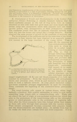
![borne in mind that Schafer observed similar formative processes in the subcutaneous connective tissue of young rats, the question must arise whether such blood-forming stations do not exist in many parts of the body and constitute seats for tlie regeneration of the blood. For purposes of demonstration it is only necessary to observe omentum of suitable agje in a fresh state in peritoneal fluid, evaporation being prevented by applying ]iaraffin to the edges of the cover-glass. Landois saw preparations of this highly interesting developmental process in the laboratory of Ranvier at Paris with such a degree of distinctness as to leave in his mind no doubt as to the accuracy of the observation. Neumann saw analogous formations in the einbrj-onal liver, Wissotzky in the amnion of the rabbit, Nicolaides in the mesen- tery of the guinea-pig, Klein in the amniotic sac of the chicken's egg, Bayerl in the cartilaginous capsules of ossifying cartilage, Leboucq and Hayem in other situations, all indicative of the fact that the blood-cells develop endogenously in certain cellular structures of considerable size whose protoplasm serves at the same time for the formation of the vessel-wall. C. -4/ a later period of life the red blood-corpuscles develop from special nucleated cells, the erythroblasts. It is believed that the latter gradually assume the form and color of perfect erythrocytes. Accord- ing to Neumann they possess blood coloring-matter from the outset. In caudate amphibia and fish the spleen, and in all other vertebrates, the bone-marrow constitutes the seat for the formation of those juve- nile forms that multiply by division. Particularly in the latter all stages of the transformation may be seen, especially pale, contractile cells resembling white blood-corpuscles, and later on red nucleated corpuscles that must be considered as the progenitors of the red corpuscles and that are capable of undergoing multiplication by mitosis. After copious loss of blood the process of transformation and the entrance into the blood-stream is said to be observed in especially marked degree. J. Arnold found in the protoplasm of the nucleated erythrocytes of bone-marrow granules resembling those of hemo- globin-free cells. In the process of transformation into red blood- corpuscles these granules become invisible through transformation. The products of the mitotic division of the pale cells especially are to be considered as the progenitors of the nucleated erythrocytes. In the red bone-marrow, perhaps also in the spleen, the small veins and most of the c&pillaries have no definite wall. The formed erythrocytes accord- ingly can at any time be swept into the circulation from these parts. The bones of the skull and most of those of the trunk contain red (blood- forming) marrow, while the extremities contain only fatty marrow, or only the upper portions of the femur and the humerus contain red inarrow. When active regenerative processes are taking place in the blood the fatty marrow may be transformed into red marrow, and indeed from the upper portion of the bones named downward, even through all the bones of the extremities. Red, blood- corpuscle-forming marrow may develop even in the ossified lar\-ngeal cartilages and in pathological bony tumors. DESTRUCTION OF RED BLOOD-CORPUSCLES. As erythrocytes are being constantly formed, it must be assumed that they are being constantly destroyed. Further, the situations are known in which this occurs especially. Among these is first the liver, as the elements of the bile are formed from blood coloring-matter and the blood of the hepatic veins contains a smaller number of red blood-corpuscles. The splenic pulp also contains cells indicative of](https://iiif.wellcomecollection.org/image/b21215418_0059.jp2/full/800%2C/0/default.jpg)