Anatomical and physiological observations on some zoophytes / by John Reid.
- John Reid
- Date:
- [1845]
Licence: Public Domain Mark
Credit: Anatomical and physiological observations on some zoophytes / by John Reid. Source: Wellcome Collection.
Provider: This material has been provided by The Royal College of Surgeons of England. The original may be consulted at The Royal College of Surgeons of England.
3/36 page 3
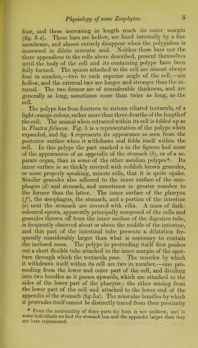
![four, and these increasing in length reach its outer margin (fig. 3 a). These bars are hollow, are lined internally by a fine membrane, and almost entirely disappear when the polypidom is immersed in dilute muriatic acid. Neither these bars nor the three appendices to the cells above described, present themselves until the body of the cell and its containing polype have been fully formed. The spines attached to the cell are almost always four in number,—two to each superior angle of the cell,—are hollow, and the external two are longer and stronger than the in- ternal. The two former are of considerable thickness, and are generally as long, sometimes more than twice as long, as the cell. The polype has from fourteen to sixteen ciliated tentacula, of a light orange-colour, rather more than three-fourths of the length of the cell. The animal when retracted within its cell is folded up as in Flustra foliacea. Fig. 5 is a representation of the polype when expanded, and fig. 4 represents its appearance as seen from the posterior surface when it withdraws and folds itself within the cell. In this polype the part marked a in the figures had more of the appearances of an appendix of the stomach (b), or of a se- parate organ, than in some of the other ascidian polypes*. Its inner surface is so thickly covered with reddish brown granules, or more properly speaking, minute cells, that it is quite opake. Similar granules also adhered to the inner surface of the oeso- phagus (d) and stomach, and sometimes in greater number to the former than the latter. The inner surface of the pharynx (/), the oesophagus, the stomach, and a portion of the intestine (c) next the stomach are covered with cilia. A mass of clark- coloured egesta, apparently principally composed of the cells and granules thrown off from the inner surface of the digestive tube, is frequently observed about or above the middle of the intestine, and this part of the intestinal tube presents a dilatation fre- quently considerably larger than what is necessary to contain the inclosed mass. The polype in protruding itself first pushes out a short flexible tube attached to the inner margin of the aper- ture through which the tentacula pass. The muscles by which it withdraws itself within its cell are two in number,—one pro- ceeding from the lower and outer part of the cell, and dividing into two bundles as it passes upwards, which are attached to the sides of the lower part of the pharynx; the other arising from the lower part of the cell and attached to the lower end of the appendix of the stomach (fig.5«). The muscular bundles by which it protrudes itself cannot be distinctly traced from their proximity * l]roiy file contractility of these parts the form is not uniform, and in some individuals we find the stomach less and the appendix larger than they are here represented.](https://iiif.wellcomecollection.org/image/b22469989_0005.jp2/full/800%2C/0/default.jpg)