Anatomical and physiological observations on some zoophytes / by John Reid.
- John Reid
- Date:
- [1845]
Licence: Public Domain Mark
Credit: Anatomical and physiological observations on some zoophytes / by John Reid. Source: Wellcome Collection.
Provider: This material has been provided by The Royal College of Surgeons of England. The original may be consulted at The Royal College of Surgeons of England.
34/36 page 16
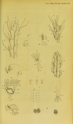
![self that in the polypes mentioned above, the inner surfaces of the polype-cells, of the appendices of those processes described in the Cellularia reptans and scruposa, of the bird-head processes, of the spines, and of the canals running along the lateral surfaces of the polypidom in Flustra avicularis, are all lined by a fine membrane. This membrane in old specimens, and when the polypes are dead, often presents numerous and pretty large cells, generally of a pale colour, at other times having a slightly yel- lowish or brownish tinge, adhering to its free surfaces. In one specimen these cells had accumulated in such quantities within some of the spines in Flustra avicularis, as to produce consider- able bulgings and excrescences. The growth and nutrition of the hard parts of the polypidom must be chiefly due to this mem- brane. EXPLANATION OF PLATE XII. Fig. 1. Magnified view of the posterior portion of the upper end of a branch of the polypidom in Cellularia reptans. It is slightly elevated on the left side, so that the polype-cells of that side are better seen than on the other. Fig. 2. Three appendices to the cells in Cellularia reptans. Fig. 3. Magnified view of four polype-cells of Cellularia reptans seen on the anterior surface. Fig. 4. Magnified view of polype in Cellularia reptans when folded up in its cell. Fig. 5. Magnified view of this polype when expanded. Fig. 6. Magnified view of the anterior surface of the upper part of one of the branches of the polypidom in Cellularia scruposa. The polype- cells are in this drawing also more distinctly seen on one side than on the other. Fig. 7. Magnified view of three appendices to the polype-cell in Cellularia scruposa ; b, b, bis, views of the process bearing the hair-like pro- longation in two different positions. Fig. 8. Greatly magnified view of head and upper part of stalk in Pedicel- lina echinata. Fig. 9. Greatly magnified view of the ciliated ova of Pedicellina echinata. Fig. 10. Magnified view of polype in Crisia chelata. Fig. 11. Magnified view of polype-cells in Alcyonidium parasiticum. Fig. 12. Magnified view of bird-head process in Flustra avicularis. Fig. 13. Magnified view of ova in Flustra avicularis. [From the Annals and Magazine of Natural History for Dec. 1845.]](https://iiif.wellcomecollection.org/image/b22469989_0036.jp2/full/800%2C/0/default.jpg)