The bronchi and pulmonary blood-vessels : their anatomy and nomenclature, with a criticism of Professor Aeby's view on the bronchial tree of mammalia and of man / by William Ewart.
- William Ewart
- Date:
- 1889
Licence: Public Domain Mark
Credit: The bronchi and pulmonary blood-vessels : their anatomy and nomenclature, with a criticism of Professor Aeby's view on the bronchial tree of mammalia and of man / by William Ewart. Source: Wellcome Collection.
Provider: This material has been provided by The University of Leeds Library. The original may be consulted at The University of Leeds Library.
41/308 page 17
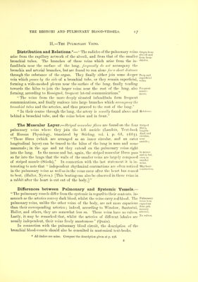
![II.—The Pulmonary Veins. Distribution and Relations.*—'• The radicles of the puhnonary veins Origin from arise from the capillary network of the alveoli, and from that of the smaller from'brou^' bronchial tubes. The branches of these veins which arise from the in- chioies. fundibula near the surface of the lung, frequently do not accompany the bronchia and arterial branches, but are found to run alone for a sliort distance through the substance of the organ. They finally either join some deeper Deep and vein which passes hy the side of a bronchial tube, or they remain superficial, veins.*' forming a wide-meshed plexus near the surface of the lung, finally tending towards the hilus to ioin the larger veins near the root of the lung, also Frequent ' tiH(isto- forming, according to Bossignol, frequent lateral communications. mosis. The veins from the more deeply situated infundibula form frequent communications, and finally coalesce into large branches which accornpany the bronchial tubes and the arteries, and thus proceed to the root of the lung. In their course through the lung, the artery is usually found above and Reintions. behind a bronchial tube, and the veins below and in front. The Muscular Layer.—Striiml mnscidar fibres are found on the four striped pulmonary veins where they join the left auricle (Landois, Text-book longit'u- of Human Physiology, translated by Stirling, vol. i. p. 68, 1885). ^^l^^.'^ These fibres (which are arranged as an inner circular, and an outer ^cav longitudinal layer) can be traced to the hilus of the lung in man and some mammals ; in the ape and rat they extend on the pulmonary veins right into the lung-. In the mouse and bat, agfain, the striped muscular fibres pass inmonse ? ^ T and 1)1 bat, SO far nito the lungs that the walls of the smaller veins are largely composed even in of striped muscle (Stieda). In connection with the last statement it is in- veius!^^ teresting to note that independent rhythmical contractions are often noticed Eliytlmiie in the pulmonary veins as well as in the vena3 cava3 after the heart has ceased ^^'^ • ■ to beat. (Haller, Nysten.) [This beating can also be observed in those veins in a rabbit after the heart is cut out of the body.] Differences between Pulmonary and Systemic Vessels.— The pulmonary vessels differ from the systemic in regard to their contents, in- asmuch as the arteries convey dark blood, whilst the veins carry red blood. The Pulmonary pulmonary veins, unlike the other veins of the body, are not more capacious capacious than their corresponding arteries ; indeed, according to Winslow, Santorini, nional'y*' Haller, and others, they are somewhat less so. These veins have no valves, aperies. Lastly, it may be remarked that, whilst the arteries of different lobules are ^° values, usually independent, their veins freely anastomose (Quain). In connection with the pulmonary blood circuit, the description of the bronchial blood-vessels should also be consulted in anatomical text-books. * All italics are mine. Compare the description given at p. 198.](https://iiif.wellcomecollection.org/image/b21518701_0041.jp2/full/800%2C/0/default.jpg)