Ellis's demonstrations of anatomy : being a guide to the knowledge of the human body by dissection / by George Viner Ellis.
- Ellis, George Viner, 1812-1900.
- Date:
- 1890
Licence: Public Domain Mark
Credit: Ellis's demonstrations of anatomy : being a guide to the knowledge of the human body by dissection / by George Viner Ellis. Source: Wellcome Collection.
35/802 (page 21)
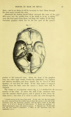
![so that the side of the face to be dissected is upwards: tix it in this position with hooks. Dissection. As a preparatory step, the muscukr fibres of the Dissection, apertures may be made slightly tense by inserting a small quantity of tow or cotton wool between the eyelids and the eyeball, and between the lips and the teeth. First lay bare the sphincter muscle of the eyelids by making a How to skin-deep incision over the margin of the orbit, and raising the skin skhrfrom of the lids towards the aperture of the eye. Much care must be eyelids, taken in detaching the skin from the thin and pale fibres of the orbicular muscle in the lids, as there is but little areolar tissue between the two. Next the integument is to be removed from the side of the face from tixe by one incision in front of the ear from above the zygomatic arch to the angle of the jaw, and another along the base of the jaw to the chin: a cut should also ])e made along the free margin of each lip from the centre to the angle of the mouth, and another round the edge of the nostril. The flap of skin is to be raised from behind forwards, and left adherent along the middle line. On the side, of the nose the skin is closely united to the subjacent and from parts, and must be detached with caution. Around the mouth is of ; the orbicular muscle of the lips, and from this many fleshy slips extend both upwards and downwards, but they are all marked so io clean distinctly as to escape injury, with the exception of the small .i-ound^ risorius muscle which goes from the corner of the mouth towards mouth, the ramus of the lower jaw. While removing the fat from the muscles, each fleshy slip may be tightened with hooks. The facial vessels and their branches will come into view as the Facial muscles are cleaned ; but the nerves may be disregarded on this side. In front of the ear is the parotid gland, the duct of which is to ai«i parotid be preserved : this is on a level with the meatus auditorius, and ^ pierces the middle of the cheek. Muscles of the Face (fig. 7). Tlie superficial muscles of the in the face face are disposed in three groups : one of the nose, another of the fOTn^three^ eyelids and eyebrow, and a third of the aperture of the mouth. One groups, of the muscles of mastication, viz., the masseter, is partly displayed at the hinder part of the face, covering the ramus of the lower jaw. Muscles of the Nose (fig. 6). These muscles are the following : Muscles of pyramidalis nasi, compressor naris, levator labii superioris alseque nasi, dilatator naris, and depressor alse nasi. The PYRAMIDALIS NASI (fig. 6,') is a small fleshy slip that covers Pyramidalis the nasal bone, and appears to be a continuation of the innermost part of the frontalis muscle. Its fibres are attached above to the skin of the forehead ; below, they end in the aponeurosis of the compressor muscles over the cartilaginous j)art of the nose. Its inner border meets the muscle of the opposite side. Action. Tliis muscle draws down the skin of the centre of the use. forehead, and produces transverse wrinkles at the root of the nose. Compressor naris. This muscle (fig. 6,^) is not well seen till Coiupressor after the examination of the following one, by which it is partly '](https://iiif.wellcomecollection.org/image/b20418358_0035.jp2/full/800%2C/0/default.jpg)