Ellis's demonstrations of anatomy : being a guide to the knowledge of the human body by dissection / by George Viner Ellis.
- Ellis, George Viner, 1812-1900.
- Date:
- 1890
Licence: Public Domain Mark
Credit: Ellis's demonstrations of anatomy : being a guide to the knowledge of the human body by dissection / by George Viner Ellis. Source: Wellcome Collection.
67/802 (page 53)
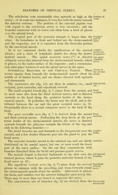
![which, will then be brought into view, contains the platysma ; and to see that muscle, it will be necessary to take the subcutaneous layer from the surface of the fibres. The PLATYSMA MYOIDES is a thin subcutaneous muscular layer, piatysma which is now seen only in its posterior half. The muscle is placed ficross the side of the neck, and extends from the top of the shoulder to the face. Its fleshy fibres take origin from the skin and sub- arises at cutaneous tissue over the clavicle and acromion, as well as from ' that covering the highest parts of the pectoral and deltoid muscles ; ascending through the neck, the fibres are inserted into the jaw and inserted the angle of the mouth. ^^^-^ ' The lower part of the muscle is more closely united to the skin than the upper, and covers the external jugular vein as well as the lower part of the posterior triangle. At first the fibres of the covers . . . triano'le' muscle are thin and scattered, but they increase in strength as they ' ascend. The oblique direction of the fibres should be noted, ])ecause in venesection in the external jugular vein the incision is to be so made as to divide them transversely. The action will be found with the description of the remainder of use. the muscle (p. 59). Dissection. The platysma is to be cut across near the clavicle. Dissection and to be reflected forwards as far as the incision over the sterno- mastoid muscle, but it is to be left attached at that spot. In raising the muscle the student must be careful of the deep fascia of the jieck ; and he should dissect out the external jugular vein, with the superficial descending branches of the cervical plexus, which are close beneath the platysma. The EXTERNAL JUGULAR VEIN (fig. 15, ^) begins just behind the External angle of the jaw by the union of the posterior division of the tem- {fn^^'^ poro-maxillary with the posterior auricular vein (fig. 16). Descend- ing beneath the platysma to the lower part of the neck, it there pierces the deep cervical fascia to open into the subclavian vein. Its course crosses side down the neck will be marked by a line from the angle of the jaw subclavian, to the middle of the clavicle. Beyond the sterno-mastoid muscle the vein is dilated, and the swollen part (sinus) is limited by two pairs of valves,—one being situate below at the mouth of the vein, and the other near the muscle. Small superficial branches join the vein, and an offset connects it with the anterior jugular vein. Its size and the height at which it crosses the sterno-mastoid muscle, are very uncertain. The DEEP CERVICAL FASCIA, like the aponeuroses in other re- Cervical gions of the body, consists of a superficial layer which surrounds the neck continuously, and of processes that are prolonged in- wards between the muscles. In some bodies this fascia is thin and indistinct. In its extent round the neck the membrane encases the sterno- mastoid, and has a different disjDOsition before and behind that muscle. As now seen passing backwards from the muscle, the Part behind fascia continues over the posterior triangular space, and encloses the ^i^astofd trapezius in its progress to the sj^ines of- the vertebrse. At the muscle](https://iiif.wellcomecollection.org/image/b20418358_0067.jp2/full/800%2C/0/default.jpg)