Ellis's demonstrations of anatomy : being a guide to the knowledge of the human body by dissection / by George Viner Ellis.
- Ellis, George Viner, 1812-1900.
- Date:
- 1890
Licence: Public Domain Mark
Credit: Ellis's demonstrations of anatomy : being a guide to the knowledge of the human body by dissection / by George Viner Ellis. Source: Wellcome Collection.
80/802 (page 66)
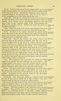
![Dissection of the subclavian artery and its brandies : of right lymphatic tiiict; of brachial plexus; of cervical plexus. Number of scalene muscles. Scalenus anticus: origin; insertion ; relations, nerves and the carotid blood-vessels. The student may examine them in the order here given. Dissection, (fig. 17). Supposing the sterno-mastoid cut, the fat and fascia are to be taken away from the lower ]3art of the neck, so as to prepare the scaleni muscles with the subclavian vessels and their branches. By means of a little dissection the anterior scalenus muscle will be seen ascending from the first rib to the lower cervical vertebrae, having the phrenic nerve and subclavian vein in front of it, the latter crossing it near the rib. The jjart of the subclavian artery on the inner side of the scalenus is then to be cleaned, care being taken not only of its branches, but also of the branches of the symj^athetic nerve which course over and along it from the neck to the chest. This dissec- tion will be facilitated by the removal of the remaining part of the clavicle. All the branches of the artery are in general easily found, except the superior intercostal, which is to be sought in the thorax in front of the neck of the first rib. On the branch (inferior thyroid) ascending to the thyroid body, or near it, is the middle cervical ganglion of the sympathetic ; and the dissector should follow down- wards from it a small cardiac nerve to the thorax. Only the origin and first part of the arterial branches can be now seen ; their termination is met with in other stages of this dissection, or in the dissection of the other parts of the body. Next the student should seek the small right lymphatic duct that opens into the subclavian vein at its junction with the internal jugular. A notice of it will be given Avith the lymphatics of the thorax. The outer part of the subclavian artery having been already prepared, let the dissector remove more completely the fibrous tissue from the nerves of the brachial plexus. From the plexus trace the small branch to the subclavius muscle, and the branches to the rhomboid and serratus muscles, which pierce the middle scalenus. If it is thought necessary, the anterior scalenus may be cut through after the artery has been studied. Clean the cervical plexus, and seek its muscular branches, the small twigs to join the descendens noni, and the roots of the phrenic nerve. Lastly, let the middle scalenus muscle be defined, as it lies beneath the cervical nerves. The SCALENI MUSCLES are usually described as three in number, and are named from their relative position, anterior, middle, and posterior ; they extend from the transverse processes of the cervical vertebrae to the first and second ribs. The SCALENUS ANTICUS (fig. 17, ^) is somewhat conical in shape, and arises from the anterior tubercles of the transverse processes of the third, fourth, fifth, and sixth cervical vertebrae. It is inserted into the upper surface and inner border of the first rib, surrounding the rough mark or projection on this part of the bone known as the scalene tubercle. More deeply seated below than above, the murtcle is concealed by](https://iiif.wellcomecollection.org/image/b20418358_0080.jp2/full/800%2C/0/default.jpg)