Ellis's demonstrations of anatomy : being a guide to the knowledge of the human body by dissection / by George Viner Ellis.
- Ellis, George Viner, 1812-1900.
- Date:
- 1890
Licence: Public Domain Mark
Credit: Ellis's demonstrations of anatomy : being a guide to the knowledge of the human body by dissection / by George Viner Ellis. Source: Wellcome Collection.
82/802 (page 68)
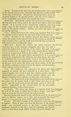
![origin insertion parts in contact with it: Scalenus posticus: attach- ments ; Subclavian artery extends to upper limb, is divided into three parts. First part, internal to scalenus, is deep. In front of, behind, and below it. Veins with the artery. antetior, and extends highest of all on the vertebrae. Its origin is from the posterior tubercles of the transverse ]:)rocesses of all the cervical vertebrae (fig. 48, d) ; and it is inserted into an impression on the upper surface of the first rib, extending from the tuberosity behind to the groove for the subclavian artery in front. In contact with the anterior surface are the subclavian artery and the cervical nerves, together with the sterno-mastoid muscle ; the posterior surface touches the posterior scalenus, and the deep lateral muscles of the back of the neck. The fibres are perforated by the nerves of the rhomboid and serratus muscles. Action. Usually it elevates the first rib. With the rib fixed, the cervical part of the spine can be inclined laterally by one muscle. The SCALENUS POSTICUS (fig. 48, f) is inconsiderable in size, and appears to be part of the preceding muscle. Arising from two or three of the lower cervical transverse processes, it is inserted below, by a thin tendon about half an inch A\dde, into the second rib in front of the serratus posticus superior. Action. It acts as an elevator of the second rib ; and its fibres having the same direction as those of the medius, it will help to incline the neck in the same Avay. The SUBCLAVIAN ARTERY (fig. 17) is the first portion of the large trunk supplying the upper limb with blood, and is thus desig- nated from its position beneath the clavicle. On the right side, this vessel (^) is derived from the bifurcation of the innominate artery behind the sterno-clavicular articulation, and the part of it named subclavian extends as far as the outer border of the first rib. To reach the limb the artery crosses the lower part of the neck, taking an arched course over the top of the lung and the first rib, and between the scaleni muscles. For the purpose of describing its numerous connections the vessel is divided into three parts ; the first extending from the sterno-clavicular articulation to the inner border of the anterior scalenus ; the second, beneath the scalenus ; and the third, from the outer border of that muscle to the outer edge of the first rib. First part. Internal to the anterior scalenus the artery lies deeply in the neck, and ascends somewhat from its origin. Between the vessel and the surface will be found the common tegumentary coverings with the platysma and the deep fascia ; the sterno- mastoid, sterno-hyoid, and sterno-thyroid muscles ; and a strong deep process of fascia froiu the inner border of the scalenus muscle. Behind and below, it rests against the pleura, which ascends into the arch formed by the vessel; and the apex of the lung separates the artery from the vertebras and the posterior ends of the first and second ribs. Veins. The innominate vein lies below and rather in front of this part of the artery. The internal jugular vein crosses the arterial trunk close to the scalenus ; and underneath this vein, with the same direction, lies the vertebral vein. Much more superficial, and separated from the artery by muscles, is the deep part of the anterior jugular vein.](https://iiif.wellcomecollection.org/image/b20418358_0082.jp2/full/800%2C/0/default.jpg)