Ellis's demonstrations of anatomy : being a guide to the knowledge of the human body by dissection / by George Viner Ellis.
- Ellis, George Viner, 1812-1900.
- Date:
- 1890
Licence: Public Domain Mark
Credit: Ellis's demonstrations of anatomy : being a guide to the knowledge of the human body by dissection / by George Viner Ellis. Source: Wellcome Collection.
88/802 (page 74)
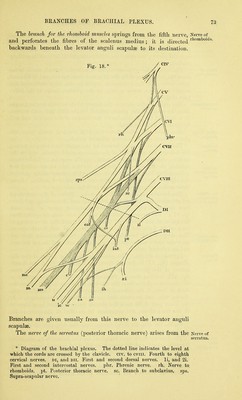
![iiftli, sixth, and generally also the seA^enth, nei^es near the inter- vertebral foramina. Piercing the fibres of the scalenus lower than the preceding branch, the nerve is continued downwards behind the brachial plexus, and enters the serratus magnus muscle on its axillary surface. The nerve oftlie suhclavius muscle is a slender branch, which arises from the trunk formed by the fifth and sixth nerves, and is directed downwards over the subclavian artery to the deep surface of the muscle ; it often sends a twig to the j)hrenic nerve at the lower part of the neck. The suprascajndar nerve is the largest of these branches, and arises from the trunk of the jjlexus formed by the fifth and sixth nerves. It is destined for the muscles on the dorsum of the scapula, and will be dissected with the arm. Occasionally an offset from the fifth cervical trunk joins the phrenic nerve on t]\e anterior scalenus muscle. The CERVICAL PLEXUS, formed by the upper four cervical nerves, lies beneath the upper half of the sterno-mastoid muscle, and on the middle scalenus and the levator anguli scapulae. Each nerve entering the plexus, except the first, divides into an ascending and a descending branch, and these unite with corresponding parts of the adjacent nerves, so as to give rise to a series of arches. From these arches or loops the different branches arise :— The branches are superficial and deep. Those of the superficial set are again subdivided into ascending and descending, and have been described with the posterior triangular space of the neck (p. 57). The ascending branches may be now seen to sj)ring from the union of the second and third nerves ; and the descending, to take origin from the loop between the third and fourth nerves. The deep set of branches remains to be examined : they are muscular and communicating, and may be arranged in an internal and an external series. Internal series. The 2)hrenic or muscular nerve of the dia- phragm (fig. 17) is derived from the fourth, or third and fourth nerves of the plexus ; and it may be joined by a fasciculus from the fifth cervical nerve. Descending obliquely on the surface of the anterior scalenus from the outer to the inner edge, it enters the chest in front of the internal mammary artery, but behind the sub- clavian vein, and traverses that cavity to reach the diaphragm. At the lower part of the neck the phrenic nerve is joined by a filament of the sympathetic, and sometimes by an off'set of the nerve to the subclavius muscle. On the left side the nerve crosses over the first jDart of tjie sub- clavian artery. The branches to the ansa hyjjoglossi are two in number : one arises from the second, and the other from the third cervical nerve. They are directed inwards over or under the internal jugular vein to join in a loop with the descending branch of the hypoglossal nerve in front of the common carotid artery (p. 77). Muscular branches are furnished to the rectus anticus major and](https://iiif.wellcomecollection.org/image/b20418358_0088.jp2/full/800%2C/0/default.jpg)