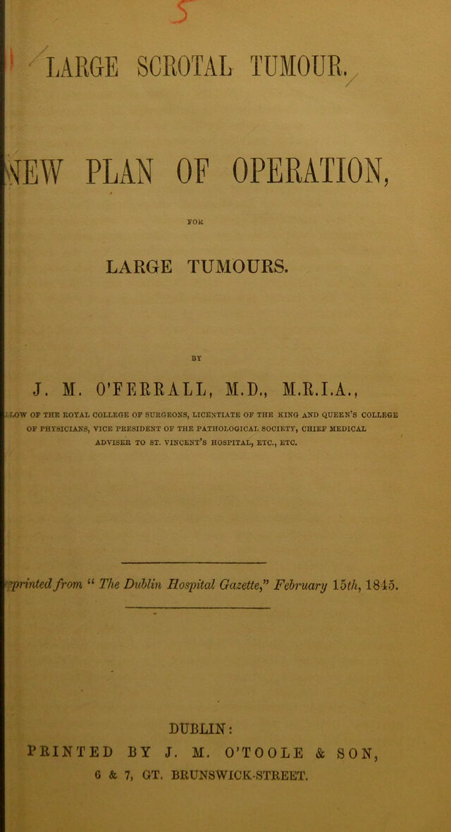
Large scrotal tumour : new plan of operation, for large tumours / by J.M. O'Ferrall.
- O'Ferrall, J. M. (Joseph Michael), 1790-1868.
- Date:
- [1845]
Licence: Public Domain Mark
Credit: Large scrotal tumour : new plan of operation, for large tumours / by J.M. O'Ferrall. Source: Wellcome Collection.
Provider: This material has been provided by The Royal College of Surgeons of England. The original may be consulted at The Royal College of Surgeons of England.
9/16 page 9
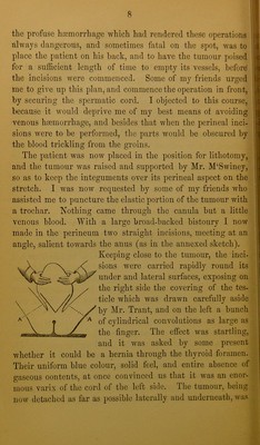
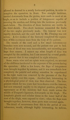
![] j Ij allowed to descend to a nearly horizontal position, in order to j; complete the operation in front. Two straight incisions, i carried downwards from the groins, were made to meet at an j 'angle, so as to include a portion of integument capable of I covering the urethra and fitting into the incisions previously I made below. The direction of these incisions are visible in the third sketch. Two short incisions connected the limbs of the two angles previously made. The tumour was noAv rapidly detached, and the cord held by Mr. Fleming was cut across. A few strokes of the bistoury completed the separa- tion of this enormous mass from the body of the patient. The vessels of the cord and a few small subcutaneous branches were now secured, and the patient was put to bed. The loss of blood was very inconsiderable, not exceeding per- haps four ounces. I cannot say what length of time was occupied by the excision, but I am assured by several gentle- men present that it was completed in eight minutes. Some warm wine and an opiate were required, on account of the chilliness incidental to the exposure of his person during the operation. After a few hours, the flaps of integument were brought together by sutures, and it was remarkable how completely the painful and aching sensation he experienced in the right testis was removed by the pressure of the flap drawn tightly over this organ. Another fact, interesting in a physiological point of view, was then observed. The inte- gument which for ten years had been drawn over the tumour, had now returned rapidly to its original position, giving the appeai-ance as if the incisions had been made across the middle of Poupart’s ligament, downwards on the thigh. The lymphatic glands and cellular tissue in the inner half of the root of the thigh, were exposed to view ; when the flaps were brought together, however, these parts were covered again. The detail of occurrences from this to the period of con- valescence, would, although highly interesting, be tedious.](https://iiif.wellcomecollection.org/image/b22345036_0009.jp2/full/800%2C/0/default.jpg)
 No text description is available for this image
 No text description is available for this image
 No text description is available for this image