The microscope: and its revelations / by William B. Carpenter; with an appendix containing the applications of the microscope to clinical medicine, etc. By Francis Gurney Smith.
- William Benjamin Carpenter
- Date:
- 1856
Licence: Public Domain Mark
Credit: The microscope: and its revelations / by William B. Carpenter; with an appendix containing the applications of the microscope to clinical medicine, etc. By Francis Gurney Smith. Source: Wellcome Collection.
585/768 page 587
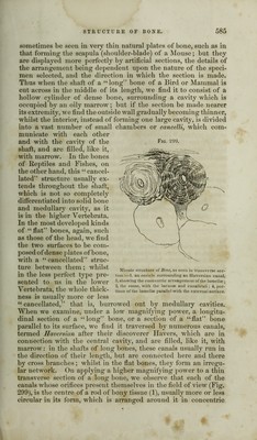
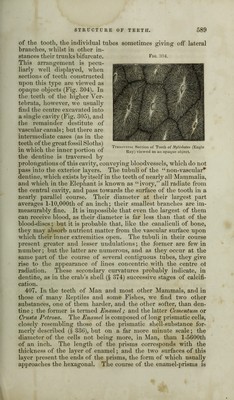
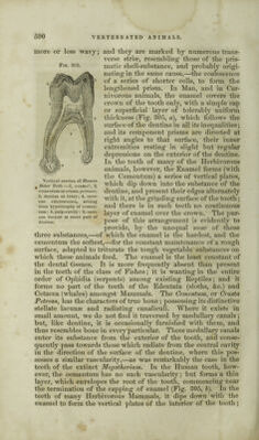
![Fig. 301. lacunae of Reptiles, without a corresponding increase in breadth ; and this is also seen in some Fishes, though in general the lacunae of the latter are remarkable for their angularity of form, and the fewness of their radiations,—as shown in Fig. 801, which repre- sents the lacunae and ca- naliculi in the bony scale of the Lepidosteus (“bony pike” of the North Ame- rican lakes and rivers), with which the hones of its internal skeleton per- fectly agree in structure. The dimensions of the lacunae in any bone do not bear any relation to the size of the animal to which it belonged; thus there is little or no per- ceptible difference between their size in the enormous extinct Iguanodon, and in the smallest Lizard now inhabiting the earth. But they bear a close relation to the size of the blood-corpuscles in the several classes; and this relation is particularly obvious in the “perennibranchiate” Batraehia, the extraordinary size of whose blood-corpuscles will be presently noticed (§ 414):— Section of the bony scale of Lepidosteus:—a. showing the regular distribution of the lacunae and of the connecting canaliculi; b, small portion more highly magnified. Proteus, Siren, Menopoma, Lepidosiren, Pterodactyle, Long Diameter. 1-570 to 1-980 1-290 to 1-480 ]-450 to 1-700 1-375 to 1-494 1-445 to 1-1185 Short Diameter. 1-885 to 1-1200 1-540 to 1-975 1-1300 to 1-2100 1-980 to 1-2200 1-4000 to 1-5225* 405. In preparing sections of bone, it is important to avoid the penetration of the Canada balsam into the interior of the lacunae and canaliculi; since, when these are filled by it, they become almost invisible. Hence it is preferable not to employ this ce- ment at all, except, it may be, in the first instance; but to rub down the section beneath the finger, guarding its surface with a slice of cork or a slip of guttapercha (§ 1 11); and to give it such a polish, that it may be seen to advantage even when mounted dry. As the polishing, however, occupies much time, the benefit which is derived from covering the surfaces of the specimen with Canada balsam may be obtained, without the injury resulting from the penetration of the balsam into its interior, by adopting the following method. A quantity of balsam proportioned to the size of the specimen is to be spread upon a glass slip, and to be * See Prof. J. Quekett’s Memoir on this subject, in the “ Transact, of the Microsc. Soc.” Ser. 1, vol. ii; and his more ample illustration of it in the “Illustrated Catalogue of the Histological Collection in the Museum of the Roy. Coll, of Surgeons,” vol. ii.](https://iiif.wellcomecollection.org/image/b28136974_0585.jp2/full/800%2C/0/default.jpg)