Illustrations of dissections : in a series of original colored plates the size of life representing the dissection of the human body / by George Viner Ellis and G.H. Ford.
- George Viner Ellis
- Date:
- 1891
Licence: Public Domain Mark
Credit: Illustrations of dissections : in a series of original colored plates the size of life representing the dissection of the human body / by George Viner Ellis and G.H. Ford. Source: Wellcome Collection.
Provider: This material has been provided by the Augustus C. Long Health Sciences Library at Columbia University and Columbia University Libraries/Information Services, through the Medical Heritage Library. The original may be consulted at the the Augustus C. Long Health Sciences Library at Columbia University and Columbia University.
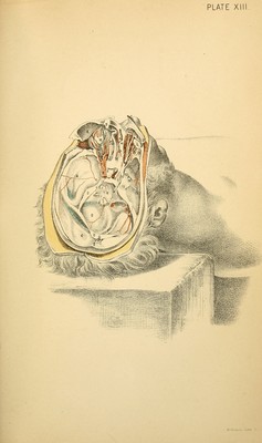
139/600 page 101
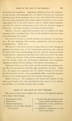
![ilie interosseous membrane. Appearing, behind, between the supinator, A., and extenso/ ossis metacar]Di, B, it is directed between the superficial and deep strata of the muscles as far as the lower third of the forearm: here it becomo-'i superficial, and courses along the tendon of the extensor carpi ulnaris, Gr, to the wrist, where it ends in offsets, which communi- cate with tJy.^ anterior interosseous,/, and with the posterior carpal, b (Plate xi.). (ts named branches are recurrent and muscular. MusciJii' branches supply the deep layer, and the digital and ulnar extensors oi the superficial layer; those to the superficial layer have been cut in <lelaching the muscles. TV recurrent branch, b, ascends between the supinator, A, and anco- neicP, H; and supplying both muscles, anastomoses with the superior proi^unda artery. (Plate vii.) The anterior interosseous artery, f, comes from the front, through an aperture in the lower part of the interosseous membrane, and ends on the back of tlie wrist, anastomosing with the posterior carpal and inter- osseous arteries; it gives a considerable offset to the outer side of the wrist. Perforating branches of the anterior interosseous arterj^, e, e, three or four in number, pierce the interosseous membrane, and anastomose together as well as with the ending of the anterior interosseous, /. Recurrent artery, g, of the radial, ascends beneath the supinator lon- gus, M, and communicates with the upper profunda in the arm. (Plate vii.) It supplies the supinator, and the radial extensors of the wrist, also the brachialis anticus; and a considerable offset enters the supinator brevis. A, and communicates with the recurrent interosseous. Radial artery, li. The anatomy of the trunk and branches of this artery on the back of the wrist and hand has been given in the descrip- tion of Plate xi., to which reference may be made. NERVE OF THE BACK OF THE FOREARM. The musculo-spiral nerve supplies the extensor and supinator muscles of the back of the forearm. 1. Musculo-spiral trunk. 2. Radial nerve. 3. Posterior interosseous. 4. Branch to the two first extensors of the tliumb. 5. Branch to the third extensor of the thumb and the indicator muscle. 6. Continuation of the posterior in- terosseous nerve. 7. Gangliform enlargement of the nerve on the wrist.](https://iiif.wellcomecollection.org/image/b2122335x_0139.jp2/full/800%2C/0/default.jpg)