Illustrations of dissections : in a series of original colored plates the size of life representing the dissection of the human body / by George Viner Ellis and G.H. Ford.
- George Viner Ellis
- Date:
- 1891
Licence: Public Domain Mark
Credit: Illustrations of dissections : in a series of original colored plates the size of life representing the dissection of the human body / by George Viner Ellis and G.H. Ford. Source: Wellcome Collection.
Provider: This material has been provided by the Augustus C. Long Health Sciences Library at Columbia University and Columbia University Libraries/Information Services, through the Medical Heritage Library. The original may be consulted at the the Augustus C. Long Health Sciences Library at Columbia University and Columbia University.
142/600 page 104
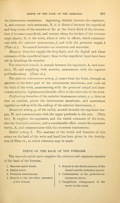
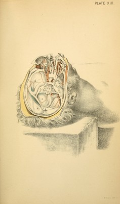
![ILLUSTRATIONS OF THE HEAD AXD ]\^ECK. DESCRIPTIOX OF PLATE XIII. The base of the skull, with the cranial nerves, and the first and second stages of the dissection of the orbit, may be studied with the aid of this Figure. After the removal of the brain, the fossae and the dura mater in the base of the skull are visible without further preparation; but the dissec- tion required for the display of the cranial nerves and the contents of the orbit will be subsequently described. BASE OF THE SKULL AND THE DURA ilATER. The region called base of the skull is situated inside the cranium, and lies below the level of a line carried circularly round the head from the superciliary eminences in front to the occipital protuberance behind. It is divided into three fossa on each side of the middle line; and a strong fibrous membrane, the dura mater, lines the whole. A. Middle fossa of the base. B. Posterior fossa. C. Superior occipital fossa. D. Part of the tentorium, cut through. E. Part of the falx cerebri, also cut. F. Falx cerebelli. G. Straight sinus. H. Cribriform plate of the ethmoid bone. I. Crista galli of the ethmoid bone. K. Roof of the orbit raised. The anterior fossa of the base lies over the orbit, and must be de- stroyed nearly altogether by the dissection of that space. For the most part the surface of the fossa is convex, but along the middle line it is hollowed where it lodges the olfactory bulb: at the forepart of the hollow, H, small apertures exist in the cribriform plate of the ethmoid bone for](https://iiif.wellcomecollection.org/image/b2122335x_0142.jp2/full/800%2C/0/default.jpg)