Illustrations of dissections : in a series of original colored plates the size of life representing the dissection of the human body / by George Viner Ellis and G.H. Ford.
- George Viner Ellis
- Date:
- 1891
Licence: Public Domain Mark
Credit: Illustrations of dissections : in a series of original colored plates the size of life representing the dissection of the human body / by George Viner Ellis and G.H. Ford. Source: Wellcome Collection.
Provider: This material has been provided by the Augustus C. Long Health Sciences Library at Columbia University and Columbia University Libraries/Information Services, through the Medical Heritage Library. The original may be consulted at the the Augustus C. Long Health Sciences Library at Columbia University and Columbia University.
39/600 page 21
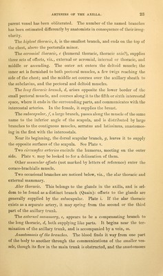
![but concealed by it, the circumflex and musculo-spiral nerves—the formei reaching- only to the edge of the subscapularis muscle. Numhcr andijosition of tlie arterial offsets. Branches are distributed internally to the thorax, and externally to the shoulder and arm. From the first part come two offsets, the highest thoracic, l, and acromial thoracic, c ; the first is small and irregular in its size and posi- tion ; and the latter, much larger, springs close to the edge of the pectoralis. Only occasionally is there any named branch on the second part. Four or five branches spring from the third part of the parent trunk. The first of these, long thoracic, d, is close to the border of the pectoralis minor. The next or subscapular branch arises opposite the loAver border of the subscapularis muscle. Two circumflex arteries take origin near the last, but they arc concealed by the trunks of the axillary vessels. The last-named branch given off is the small external mammary, e. Ligature of the artery.—The axillary artery may be tied near the clavicle, as ■well as near the ending (p. G). Near the clavicle, or above the small pectoral muscle, the vessel lies deeply, and is reached only after cutting through the pectoralis major. Two offsets, superior and acromial thoracic, spring usually from this part of the artery, wath the supra-scapular (a branch of the subclavian) some- times, and they leave scarcely interval enough for the application of a ligature, especially if the first is large. The connections also of the artery with superficial vessels and nerves are so complicated (see Plate) as to render hazardous ligature of it at this s]3ot. The vessel might be tied in this situation for aneurism of the lower 2?art of the arterial trunk, or for the arrest of hemorrhage after an opera- tion high up the arm ; but the difficulties in securing the vessel, and the chances of recurring bleeding, may almost deter a surgeon from having recourse to the operation. Should it be necessary to ligature the artery here, a practical knowl- edge of the anatomy will assist the operator in his attem2:)ts to secure the vessel. With the arm outstretched, the position of the artery Avill be marked by a line over the surface of the pectoralis major, which has been described already (p. 20). Tho surface depressions on the sides of the clavicular attachment of the pectoralis major being taken as the limit of the incisions, the operator](https://iiif.wellcomecollection.org/image/b2122335x_0039.jp2/full/800%2C/0/default.jpg)