Diseases of the heart and aorta / by Thomas E. Satterthwaite.
- Satterthwaite, Thomas E. (Thomas Edward), 1843-1934
- Date:
- [1905]
Licence: Public Domain Mark
Credit: Diseases of the heart and aorta / by Thomas E. Satterthwaite. Source: Wellcome Collection.
Provider: This material has been provided by the Augustus C. Long Health Sciences Library at Columbia University and Columbia University Libraries/Information Services, through the Medical Heritage Library. The original may be consulted at the the Augustus C. Long Health Sciences Library at Columbia University and Columbia University.
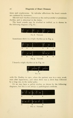
22/320 page 18
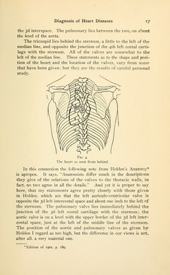
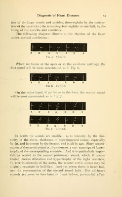
![Seen from behind, the npijcr level of the heart corresponds •\\ ith the center of the 4th dorsal vertebra, and the lower margin of rtlie 5tli rib, on the left side; the ni:)per margin of the 6th rib on the right side. The apex is opposite the Sth left interspace, abont midway between the spines of the vertebra; ami the free border of the ribs. The mitral valve is opposite the 6th interspace, close to the left margin of the 6th dorsal vertebra. The aortic lies to the left of the median line, opposite the point where the 5th dorsal «pine overlaps the 6th ; the pulmonary lies between them ; the tricus- ]iid covers the median line, though slightly more to the left than the right, and is opposite the root of the spine of the 6th dot sal verte- t.ra (Fig. 4). In considering heart murmurs the physiological action of the 'heart must be taken into consideration. The movement of the 'blood is caused by the contraction of the auricles, ventricles and vessels. The blood enters the auricles by the veins, and then is ■ex])elled by the auricles through the auriculo-ventricular open- ings or valves into the ventricles; when the ventricles are filled, they contract and force this blood back into the vessels, the left ventricle driving a column of blood through the aorta into the .greater or systemic circulation : the right \entricle driving an- •other column of blood into the lesser or pulmonary circulation. 'Then follows a contraction of the great vessels, the aorta and pulmonary artery. In health the action of the ventricles in clos- ing is attended with a sound or tone, due to three principal causes; i. The closure of the auricular-ventricular orifices. 2. The muscular action of the ventricles. 3. The vibration of blood in the ventricles. Roth auricles and great vessels contract during the filling of the ventricles (diastole) and hence any sound produced during this period is called diastolic, but they are not synchronous, the vessels contracting at the beginning of diastole, and the auricles at the end. A systolic sound is produced during the contraction of the ventricles (systole), and the word presystolic is accepted as indicating a sound produced at the end of diastole, or during the contraction of the auricles: or in fact, any sound not produced during the time for the contraction of the aorta. The second sound is chiefly due to the closure of the aortic and pulmonary valves, and the vibration of blood in the aortic and pulmonary arteries. Valves, muscular action, chordse tendinge, vessels and the vibration of the blood, produce heart sounds. Now, supposing the time occupied by these actions were di- • vided into eighths, one-eight would be occupied by the contrac-](https://iiif.wellcomecollection.org/image/b21208384_0022.jp2/full/800%2C/0/default.jpg)