The Hunterian lectures on the pathology and surgery of intussusception : delivered at the Royal College of Surgeons of England / by D'Arcy Power.
- Power, D'Arcy, 1855-1941.
- Date:
- 1897
Licence: Public Domain Mark
Credit: The Hunterian lectures on the pathology and surgery of intussusception : delivered at the Royal College of Surgeons of England / by D'Arcy Power. Source: Wellcome Collection.
Provider: This material has been provided by The Royal College of Surgeons of England. The original may be consulted at The Royal College of Surgeons of England.
3/38
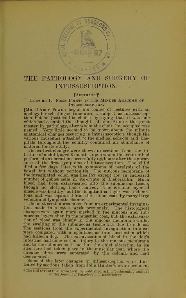
![THE PATHOLOGY AND SURGERY OF INTUSSUSCEPTION. [Abstract.]1 Lecture I.—Some Points in the Minute Anatomy op Intussusception. [Mr. D'Arcy Power began his course of lectures with an apology for selecting so time-worn a subject as intussuscep- tion, but he justified his choice by saying that it was one which had occupied the thoughts of John Hunter, the great master in pathology, after whom the chair he occupied was named. Very little seemed to be known about the minute anatomical changes occurring in intussusception, though the various museums attached to the medical schools and hos- pitals throughout the country contained an abundance of material for its study. The earliest changes were shown in sections from the in- testine of a child, aged 8 months, upon whom the lecturer had performed an operation successfully 13^ hours after the appear- ance of the first symptoms of intussusception. The child died a few days later, with symptoms of paralysis of the bowel, but without peritonitis. The mucous membrane of the invaginated colon was healthy except for an increased number of goblet cells in its crypts of Lieberkiihn. Much blood had been extravasated into the submucous tissue, though no clotting had occurred. The circular layer of muscle was healthy, but the longitudinal layer was cedema- tous, and was separated from the serous coat by many large venous and lymphatic channels. The next section was taken from an experimental invagina- tion made in a cat a week previously. The histological changes were again more marked in the mucous and sub- mucous layers than in the muscular coat, but the extravasa- tion of blood was chiefly in the mucous membrane whilst the swelling of the submucous tissue was due to oedema. The sections from the experimental invagination in a cat were compared with a spontaneous intussusception which had killed a dog. The extravasation of blood in the dog's intestine had done serious injury to the mucous membrane and to the submucous tissue, but the chief alteration in its structure had taken place in the muscular coat, where the circular fibres were separated by the oedema and had degenerated. Some of the later changes in intussusception were illus- trated by sections taken from John Hunter's own specimen, 1 The full text of this lecture will be published in the forthcoming number of the Journal of Pathology and BacteHology.](https://iiif.wellcomecollection.org/image/b22322784_0005.jp2/full/800%2C/0/default.jpg)