A series of engravings, accompanied with explanations, which are intended to illustrate the morbid anatomy of some of the most important parts of the human body. Divided into ten fasciculi / [Matthew Baillie].
- Matthew Baillie
- Date:
- 1812
Licence: Public Domain Mark
Credit: A series of engravings, accompanied with explanations, which are intended to illustrate the morbid anatomy of some of the most important parts of the human body. Divided into ten fasciculi / [Matthew Baillie]. Source: Wellcome Collection.
31/394 (page 19)
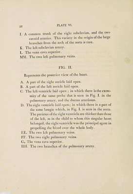
![\ [ 19 ] PLATE V. The object of this Plate is to represent a large ossification upon the surface of the heart. Ossification of the pericar¬ dium, or of the muscular structure of the heart, occurs very rarely, although ossification of the valves, more especially at the origin of the aorta, is not uncommon. A. A part of the right ventricle, just at the origin of the pul¬ monary artery. B. A part of the ascending aorta, as it is about to form the arch. C. The common trunk of the right carotid and right subcla¬ vian arteries. D. The left carotid artery. E. The left subclavian artery. HG. Two branches of the right branch of the pulmonary artery. FF. Part of the descending aorta, which is smaller than the natural size. This had been occasioned by the blood being propelled into it from the left ventricle, in less than the natural quantity, and by a feeble exertion I. The vena cava superior, somewhat enlarged beyond the natural size, K, The vena cava inferior.](https://iiif.wellcomecollection.org/image/b3045301x_0031.jp2/full/800%2C/0/default.jpg)