Anatomy, descriptive and surgical / by Henry Gray ; the drawings by H.V. Carter ; with additional drawings in later editions.
- Henry Gray
- Date:
- 1893
Licence: Public Domain Mark
Credit: Anatomy, descriptive and surgical / by Henry Gray ; the drawings by H.V. Carter ; with additional drawings in later editions. Source: Wellcome Collection.
64/1178 page 18
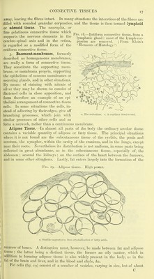
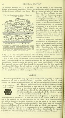
![the average diameter of of an inch. They are formed of an exceedingly delicate protoplasmic membrane, filled with fatty matter, which is liquid during life, but becomes solidified after death. They are round or spherical where they have not been subjected to pres- FiG. 20.—Development of fat. (Klein and sure ; otherwise they assume a more Noble Smith.) or less angular outline. A nucleus is always present and can be easily demonstrated by staining with log- wood ; in the natural condition it is so compressed by the contained oily matter as to be scarcely recog- nisable. These fat-cells are con- tained in clusters in the areolae of fine connective tissue, and are held together mainly by a network of capillary blood-vessels, Avhich are distributed to them. Fat is an inorganised substance, consisting of a liquid material (gly- cerine) in combination with certain fatty acids, stearic, palmitic, and oleic. Sometimes the acids sepa- rate spontaneously before the fat is examined, and are seen under the microscope in a crystalline form, as in fig 19, a. By boiling the tissue in ether or strong alcohol, the fat may be extracted from the vesicle, which is then seen empty and shrunken. Fat is said to be first detected in the human embryo about the fourteenth week. According to Klein, the fat-cells are formed by the transformation of the protoplasmic connective-tissue corpuscles, into which small globules of fat find their way and increase until they distend the corpuscle into the thin mantle of protoplasm which forms the cell-wall, and in which its nucleus is still to be seen (fig. 20). Others of the connective-tissue corpuscles are transformed into the vessels and the lymphatic tissue which accompanies the vessels. . Minute artery, v. Aliuutevem. c. Capillary bluod-vessels in the course ot tormation ; they are not yet completely hollowed out, there being stiii lelt in them protoplasmic septa, d. The ground-substance, containing numerous nucleated cells, some of which are more distinctly branched and flattened than others, and appear therefore more spindle-shaped. PIGMENT In various parts of the body ijigvient is found ; most frequently in epithelial cells and in the cells of connective tissue. Pigmented epithelial cells are found forming the external layer of the retina (fig. 21) and on the posterior surface of the iris. Pigment is also found in the epithelial cells of the deeper layers of the cuticle in some parts of the body—such as the areola of the nipple and in coloured patches of skin and especially in the skin of the coloured races, and also in hair. ^^^^ It is also found in the epithelial cells of the olfactory region, 1^^^ and of the membranous labyrinth of the ear. In the connective-tissue cells pigment is frequently met with in the lower vertebrates. In man it is found in the choroid coat of the eye, and in the iris of all but the light blue eyes and the albino. It is also occasionally met with in the cells of retiform tissue and in the pia mater of the upper part of the spinal cord. These cells are characterised by their larger size and branched processes, which, as well as the body of the cells, are filled with granules. The j pigment consists of dark brown or black granules of very small size closely packed] together within the cells, but not invading the nucleus. Occasionally the pigment-, is yellow, and when occurring in the cells of the cuticle constitutes ' freckles.'](https://iiif.wellcomecollection.org/image/b20402934_0064.jp2/full/800%2C/0/default.jpg)