The organs of vision : their structure and functions / by G.J. Witkowski ; tr. by Henry Power.
- Gustave-Joseph Witkowski
- Date:
- [1878?]
Licence: Public Domain Mark
Credit: The organs of vision : their structure and functions / by G.J. Witkowski ; tr. by Henry Power. Source: Wellcome Collection.
Provider: This material has been provided by The Royal College of Surgeons of England. The original may be consulted at The Royal College of Surgeons of England.
38/44 (page 38)
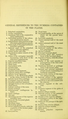
![GENEEAL EEFERENCES TO THE NUMBERS CONTAINED IN THE PLATES. 1. Palpebral conjunctiva. 2. Ocular conjunctiva. 3. Semilunar fold of the conjunctiva (plica semilunaris). 4. Periorbital portion of the orbicu- laris palpebrarum muscle. 5. ' Palpebral and ciliary portion of the orbicularis palpebrarum muscle. 6. Outer angle of the palpebral fissure (external canthus). 7. Internal angle of the palpebral fissure (internal canthus). 8. Ciliary border and cilia. 9. Zygomaticus minor muscle of the left side. 10. Fibres of the corrngator supercilii decussating 'with those of the orbicularis palpebrarum. 11. Superior maxillary bone. 12. Infra orbital nerve. 13. Malar bone. 14. Supraciliary arch. 15. Frontal bone. 16. Temporal fossa. 17. Superior broad ligament. 18. Superior tarsal fibro-cartilage. 19. Inferior broad ligament. 20. Orifices of the Meibomian glands on the ciliary border of the lid. 21. Inferior tarsal fibro-cartilage. 22. Corrugator supercilii muscle. 23. External palpebral ligament. 24. Straight tendon of the orbicularis. 25. Nasal bones. 26. Lateral cartilage of the nose. 27. Septal cartilage. 28. Supplemental cartilaginous la- mellae. 29. Nasal cartilage. 30. Ala nasi. 31. Ascending process of the superior maxillary bone. .32. Fossa for the lacrymal gland. 33. Nasal fossae. 34. Maxillary sinus. 35. Meibomian glands. 36. Orbito-palpebral muscle, with the expansion of the levator palpe- bne superioris. 37. Lacrymal sac. 38. Nasal duct. 39. Lacrymal papillae, at the apices of which are the puncta lacry- malia. 40. Lacrymal canaliculi. 41. Antero-internal wall of the nasal duct. 42. Antero-external wall of the nasal duct. 43. Caruncula lacrymalis. 44. Refiected tendon of the orbicu- laris palpebrarum. 45. Horner’s muscle. 46. Principal lacrymal gland. 47. Accessory gland and excretory ducts of the lacrymal gland. 48. Capsule of Tenon, with the orbital aponeurosis. 49. Fasciae of the recti muscles or prolongations of the first order of the capsule. 50. Tendinous fasciculi or prolonga- tions of the second order. 51. Internal and external unstriated orbital muscle terminating in a tendinous expansion. 52. Fascia of the tendon of the supe- rior oblique muscle. 53. Internal surface of the anterior segment of the capsule of Tenon. 54. ] nsertions of the recti muscles into the sclerotic. 55. Anterior segment of the scle- rotic. 56. Iris. 57. Posterior segment of the globe of the eye. 58. Inferior oblique muscle. 59. Internal surface of the posterior segment of the capsule. 60. Tendon of the superior oblique. 61. Cartilaginous pulley of the supe- rior oblique. 62. Orifice for the passage of the optic nerve. 63. Fascia of the inferior oblique. 64. Superior oblique muscle. 65. Optic nerve. 66. Levator palpebrfe superioris muscle.](https://iiif.wellcomecollection.org/image/b22465637_0040.jp2/full/800%2C/0/default.jpg)