Volume 1
Operative gynecology / by Howard A. Kelly ; with ... illustrations for the most part by Max Brödel.
- Howard Atwood Kelly
- Date:
- 1906
Licence: Public Domain Mark
Credit: Operative gynecology / by Howard A. Kelly ; with ... illustrations for the most part by Max Brödel. Source: Wellcome Collection.
590/724 (page 558)
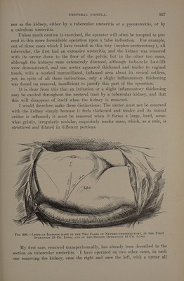
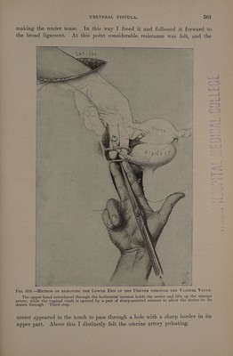
![the way down to the vesical end. In the second case I lengthened the lumbar incision down to a point just above the pubic spine, and by detaching the peri- toneum from the iliac fossa and the lateral pelvic wall, succeeded in taking out the right ureter, after doubly ligating and cutting the uterine vessels, without tying any other vessels or opening the peritoneum at any point (see Fig. 306). Removal of the Right Kidney and Ureter through a Short Lumbar and a Vaginal Incision (see Johns Hopkins [Hostal Bul- letin, Feb., 1896, p. 34).—The plan of opera- tion adopted in this case worked so well that I shall describe it fully. The. patient (CK. W.; J. H.:H., No.-4012, Dec. 21, 1895) was a large, stout woman, weighing 225 pounds, and thirty years old. She had suffered for two years with attacks of violent pain, beginning in the region of the right kidney and extending around to the front of the abdomen and down into the pelvis.. She also suffered from frequent, burning micturi- tion. There was some pus in the urine, but she had never passed any blood or a stone. The attacks of pain, which at first were in- frequent, finally came on as often as three or Hie. 307.-Removat or Kip four times weekly, beginning under the right (Nepuro-ureTeREctoMY). shoulder blade. They were so violent that she A a payee was wont to throw herself down on the floor two lower pieces of the ureter sereaming. below the ligature were re- 5 2 moved through the vaginal A urinary analysis, made after catheteriz- vault. 4 natural size (see. ] Figs. 308-311). W. Op. Dxc. ing both ureters, showed that the urine from mee the right side contained pus while that from the left was free from it, and that the percentage of urea from the right kidney was 2.1, while it was 2.6 from the left kidney. No tubercle bacilli could be found. O peration.—The fat on the abdominal walls was 7 cm. thick, and the margin of the ribs close to the crest of the ilium. __ A transverse incision was made, beginning in front of the quadrate lumbar muscles and extending 16 em. across the abdomen in the umbilical line, reaching almost to the right linea semilunaris. Numerous bleeding vessels were clamped and tied with catgut. The first lumbar nerve, with vessels accompanying it, was divided between the transversalis and the peri- toneum in the posterior part of the wound. The perirenal fat was freed on all sides of the kidney, completely detached, and brought out of the incision. By drawing it down over the lower lip of the incision the renal vessels were exposed, with the pelvis of the kidney lying beneath them.](https://iiif.wellcomecollection.org/image/b32841863_0001_0590.jp2/full/800%2C/0/default.jpg)