Lectures on surgical pathology, delivered at the Royal College of Surgeons of England / by James Paget.
- James Paget
- Date:
- 1863
Licence: Public Domain Mark
Credit: Lectures on surgical pathology, delivered at the Royal College of Surgeons of England / by James Paget. Source: Wellcome Collection.
844/880 (page 816)
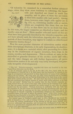
!['If tubercles be examined in a somewhat further advanced stage, when they show more tendency to softening, the larger Fig. 117* cells just described are found in much less 5' quantity, and in place of them the air-vesicle ^tl^,® ft filled with smaller cells [and nuclei]. Among /4 °^^°|€®''!^Sf however, some larger cells appear (as in ^SfWf fig- 117, a), containing smaller cells or nuclei, * ® ^ which are completely like those that are free %W (& 6); so that there can be no doubt but that, in this state, the larger corpuscles are dissolved or burst; and the smaller ones set free.' These smaller cells and nuclei set free are what have been generally described as the tubercle corpuscles; and, as I have abeady said, the tuberculous deposits,, after the earliest periods of their formation, may appear to contain no other formed corpuscles besides them.f Now the most peculiar character of tuberculous matter which these descriptions illustrate, is its early degeneration, its abortive- ness; it is shown as a material which, after proceeding for a little way in the acquirement of organic structure, then stops in its course, recedes, and degenerates. This is evident, at once, in the shrivelled or granular structure of the set-free nuclei and cells ; and the later changes are still further degenerative; all prove tuberculous matter to be not only very lowly developed, but gene- rally incapable of development-^ * Fig. 117. Tubercle-corpuscles: magnified 420 times and described in the text. Copied from SeiLToeder van der Kolk. t In an elaborate paper in the Br. & For. Med. Chi. Eev., April 1855, On the Development of Tubercle, Dr. C. E. Hall states that the formation of tubercle in the lungs is accompanied and preceded by fatty degeneration of many of the epithelium cells of the air-vesicles. He looks upon the large, many nucleated cells as modified epithelium cells, but does not, as Van der Kolk seems to do, limit them to the central parts of the cavities of the air-cells. He states that they may be found of the largest size and containing their largest number of nuclei, while closely adherent to the wall of the air-vesicle. Neither does he consider that the fi-ee tubercle corpuscles are solely derived from the nuclei of the large cells, set free by the bursting of the walls, but that the proper tubercle corpuscles are mostly formed in and from the plasma exuded into the air-vesicles. Virchow, however, in conformity with his views that the corpuscles of pus and cancer are developed from the pre-existing cells and nuclei of the textures, has, by carrying out the same methods of investigation, traced the development of tubercle corpuscles to proliferating changes in the corpuscles of^ tlie connective-tissue of the part in which the tubercle iirises. (Cell. Path. Lecture XX.) Fiirster also has illustrated in his Atlas (Taf. xxxvi. fig. 1) the development of tubercle from the connective-tissue corpuscles in padmonary tuberculosis. In a recent paper in Vii-chow's Ai-chiv (xxiv. p. 671), Eindfleisch has traced the mode of formation of the cells of miliary tubercle in a case of acute hydrocephalus. He found the various ramifications of the arteries of the pia mater studded with numerous millet-seed like granules, and he traced the development of the cells, of which these granules wore composed, from the nuclei of the external coat of the arteries to which they were connected. I An exception to this statement must be made, for certain cases, in which one part](https://iiif.wellcomecollection.org/image/b20397550_0850.jp2/full/800%2C/0/default.jpg)