Dr. J. Burdon Sanderson's reports of an experimental study of infective inflammations.
- Burdon-Sanderson, John Scott, 1828-1905.
- Date:
- [1875]
Licence: Public Domain Mark
Credit: Dr. J. Burdon Sanderson's reports of an experimental study of infective inflammations. Source: Wellcome Collection.
Provider: This material has been provided by The Royal College of Surgeons of England. The original may be consulted at The Royal College of Surgeons of England.
8/34 page 54
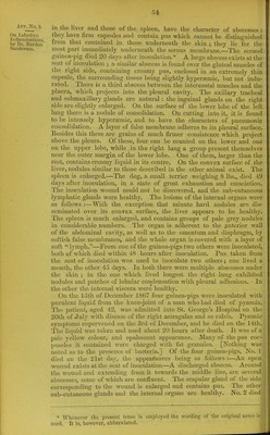
![Arr. No. 2. On Infectivo Inflammations, by Dr, bunion Sanderson. in the liver mid those oi the spleen, have the character of abscesses : they have firm capsules and contain pus which cannot be distinguished from that contained in those underneath the skin ; they lie for the most part immediately underneath the serous membrane.—The second guinea-pig died 20 days after inoculation.* A large abscess exists at the seat of inoculation ; a similar abscess is found over the gluteal muscles of the right side, containing creamy pus, enclosed in an extremely thin capsule, the surrounding tissue being slightly hyperaemic, but not indu- rated. There is a third abscess between the intercostal muscles and the pluera, which projects into the pleural cavity. The axillary tracheal and submaxillary glands arc natural: the inguinal glands on the right side are slightly enlarged. On the surface of the lower lobe of the left lung there is a nodule of consolidation. On cutting into it, it is found to be intensely hyperaemic, and to have the characters of pneumonic consolidation. A layer of false membrane adheres to its pleural surface. Besides this there are grains of much firmer consistence which project above the pleura. Of these, four can be counted on the lower and one on the upper lobe, while .in the right lung a group present themselves near the outer margin of the lower lobe. One of them, larger than the rest, contains creamy liquid in its centre. On the convex surface of the liver, nodules similar to those described in the other animal exist. The spleen is enlarged.—The dog, a small terrier weighing 8 lbs., died 49 days after inoculation, in a state of great exhaustion and emaciation. The inoculation wound could not be discovered, and the sub-cutaneous lymphatic glands were healthy. The lesions of the internal organs were as follows :—With the exception that minute hard nodules are dis- seminated over its convex surface, the liver appears to be healthy. The spleen is much enlarged, and contains groups of pale grey nodules in considerable numbers. The organ is adherent to the anterior wall of the abdominal cavity, as well as to the omentum and diaphragm, by softisli false membranes, and the whole organ is covered with a layer of soft “ lymph.”—From one of the guinea-pigs two others were inoculated, both of which died within 48 hours after inoculation. Pus taken from the seat of inoculation was used to inoculate two others; one lived a month, the other 45 days. In both there were multiple abscesses under the skin ; in the one which lived longest the right lung exhibited nodules and patches of lobular condensation with pleural adhesions. In the other the internal viscera were healthy. On the loth of December 1867 four guinea-pigs were inoculated with purulent liquid from the knee-joint of a man who had died of pyaemia. The patient, aged 42, was admitted into St. George’s Hospital on the 20th of July with disease of the right astragalus and os calcis. Pytemic symptoms supervened on the 3rd of December, and he died on the 14tli. The liquid was taken and used about 20 hours after death. It was of a pale yellow colour, and opalescent appearance. Many of the pus cor- puscles it contained were charged with fat granules. [Nothing was • noted as to the presence of bacteria.] Of the four guinea-pigs, No. 1 died on the 21st day, the appearances being as follows :—An open j wound exists at the seat of inoculation—A discharged abscess. Around , the wound and extending from it towards the middle line, are several ! abscesses, some of which are confluent. The scapular gland of the side I corresponding to the wound is enlarged and contains pus. The other I sub-cutaneous glands and the internal organs are healthy. No. 2 died j * Whenever the present tense is employed the wording of the original notes is J used. It is, however, abbreviated.](https://iiif.wellcomecollection.org/image/b22356873_0010.jp2/full/800%2C/0/default.jpg)


 No text description is available for this image
 No text description is available for this image
 No text description is available for this image