A treatise on the diseases of the eye / By J. Soelberg Wells... Together with selections from the test-types of Prof. E. Jaeger and Prof. H. Snellen.
- Wells, J. Soelberg (John Soelberg), -1879
- Date:
- 1883
Licence: Public Domain Mark
Credit: A treatise on the diseases of the eye / By J. Soelberg Wells... Together with selections from the test-types of Prof. E. Jaeger and Prof. H. Snellen. Source: Wellcome Collection.
Provider: This material has been provided by the Harvey Cushing/John Hay Whitney Medical Library at Yale University, through the Medical Heritage Library. The original may be consulted at the Harvey Cushing/John Hay Whitney Medical Library at Yale University.
87/898 page 93
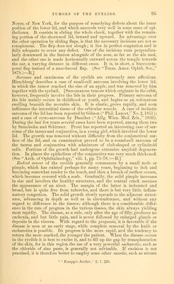
![Another remarkable case is reported by Dr. Henry Shaw/ of the Massa- chusetts Eye aud Ear Infirmary. The man was fifty-six years of age, and the horn, which was situated on tlie right lower lid, attained a length of one and three-fourths inch, its circumference at its base being one and seven- eighths inch ; it was curved, and looked like the beak of a bird. Dr. Shaw excised it with success. £pithelial cancer is almost the only malignant tumor which occurs pri- marily in the eyelids, for the other forms, such as scirrhus, medullary cancer, etc., are generally only secondarily met with in this situation. Epithelial cancer shows itself most frequently in the lower eyelid, and near the outer canthus. [It is of three kinds: the superficial, the deep, and the papillomatous; and it is said that these are all different stages of the same disease.—B.] It occurs generally in persons above the age of forty, or even in those much more aged, being rarely met with in youthful individuals. At the outset, the disease assumes the appearance of a small, circumscribed, slightly elevated induration, situated at, or close to, the edge of the lid, and looking like a wart or a small thickened crust. It is covered by healthy- looking, uniuflamed skin, and a few varicose vessels are perhajDs seen to pass over or near it. The surface of the little nodule often looks rough and scaly, as if the cuticle were thickened. It may remain in this condition for a very Jong period, and years may elapse before it increases materially in size, or becomes ulcerated. On this account, and from its being quite painless, it is often entirely disregarded by the patient, who supposes it to be simply a wart. When the disease occurs in the skin over the lachrymal sac, it has been mistaken for dacryocystitis. Thus Mackenzie mentions one instance, in which the patient called to have a style introduced, and another, in which one had actually been worn. But, sooner or later, it gradually and almost imperceptibly increases somewhat in size, creeping along the edge of the lid and assuming a lengthened, ovoid shape. Its surface becomes broken and excoriated, and a thin grayish-yellow discharge exudes from it, which hardens upon it in the form of dark rough crusts. Then ulceration sets in, and the tumor slowly spreads in circumference and depth, the edges of the ulcer being somewhat elevated, and studded, perhaps, with a few palish-red tuber- cles, which rapidly form again if abscised. The skin around the tumor is but little thickened, swollen, or discolored, and this distinguishes the disease from lupus, and also from a syphilitic ulcer. Moreover, the slowness of its growth and the history of the case, would prevent its being mistaken for the latter. When the ulceration sets in, the pain increases, but seldom to any considerable degree, nor is it of a very acute, lancinating character; but if any nerves are exposed by the ulceration, the patient's suffering will, of course, be much augmented. The discharge is of a yellowish color, healthy in nature, and free from fetor.^ Sometimes, the ulcer may become tempo- rarily cicatrized, either completely or in part, and then remain apparently healed for a certain time; but soon a breach of surface again occurs, and fresh ulceration sets in. In time, the ulcer invades the lid more and more, spreading along its surface and extending deeply into its structure, until it may eat its way completely through its whole thickness, and appear on the conjunctival surface; thence, perhaps, extending to the orbit. If the lids are destroyed, the eyeball will be exposed, and suppuration of the cornea may ensue, accompanied perhaps by loss of the lens and a considerable por- tion of the vitreous humor, and followed by atrophy of the globe. Mac- 1 Boston Med. and Surg. .Journal, 1869, Feb. 11. ^ Vide Dr. Jacob's able paper on this disease, Dublin Hospital Keports, vol. iv., 1827.](https://iiif.wellcomecollection.org/image/b20999392_0087.jp2/full/800%2C/0/default.jpg)
 No text description is available for this image
 No text description is available for this image
 No text description is available for this image