Licence: Public Domain Mark
Credit: Surgery of the ureter / by Christian Fenger. Source: Wellcome Collection.
Provider: This material has been provided by The Royal College of Surgeons of England. The original may be consulted at The Royal College of Surgeons of England.
21/42 (page 19)
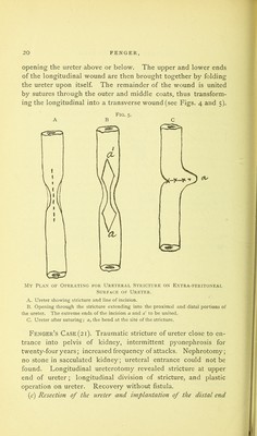
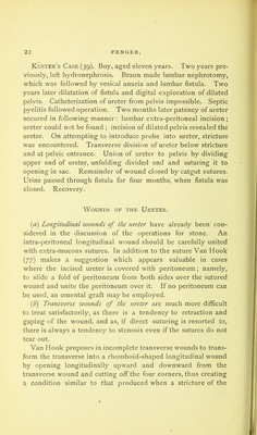
![Fig. 4. Illustrating Operation for Stricture of Ureter. A. Sacculated kidney, dilated pelvis, ureter with stricture at its upper end. i, kid- ney,- 2, sacs corresponding to dilated calices; 3, nephrotomy; 4, dilated pelvis; 5, opening in posterior surface of pelvis, pelviotomy wound; 6, ureter below stricture; 7, stricture in upper end of ureter; 8, opening in ureter below stricture, extending up through the stricture into the pelvis; 9, sutures closing the upper half of the wound in the pelvis ; a, a' and b, b', points of incision in ureter and pelvis to be united by sutures after folding the ureter upon itself at the place of stricture. B. Pelvis and ureter after union by sutures, i, pelvis; 2, ureter; 3, fold of ureter at place of stricture ; 4, sutures of wound in pelvis; 5, place of sutures between points a, a' and b and b'] 6, additional sutures, as many as needed, to close the borders of the fold formed by approximation o a io a' and b to b'.](https://iiif.wellcomecollection.org/image/b22453374_0023.jp2/full/800%2C/0/default.jpg)