An examination of phrenology; in two lectures, delivered to the students of the Columbian College, District of Columbia, February, 1837 / By Thomas Sewall.
- Thomas Sewall
- Date:
- 1838
Licence: Public Domain Mark
Credit: An examination of phrenology; in two lectures, delivered to the students of the Columbian College, District of Columbia, February, 1837 / By Thomas Sewall. Source: Wellcome Collection.
7/98
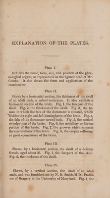
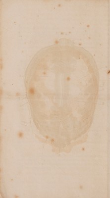
![EXPLANATION: OF THE PLATES. Plate I. Exhibits the name, form, size, and position of the phre- nological organs, as represented on the figured head of Mr. Combe. It also shews the form and application of the - craniometer. Plate IT. : Shews by a horizontal section, the thickness. of the skull of an adult male, a robust waterman. It also exhibitsa horizontal section of the brain, Fig. 1. the forepart of the skull. Fig. 2, the thickness of the skull. Fig. 3, the fis- sure in which the falx of the duramater is situated, which “divides the right and left hemispheres of the brain. Fig. 4, the falx of the duramater turned back. Fig. 5, the cortical or pulpy part of the brain. Fig. 6, the medullary or fibrous portion of the brain. Fig. 7, the grooves which separate the convolutions of the brain. Fig. 8, the corpus callosum, or great commissure of the brain. Plate III. Shews, by a horizontal section, the skull of a delicate female, aged about 25. Fig. 1, the forepart of the skull. Fig. 2, the thickness of the skull. Plate IV. Shews, by a vertical section, the skull of an adult male, and was furnished me by N. R. Smith, M.D., Profes- sor of Surgery in the University of Maryland, Fig. ], the](https://iiif.wellcomecollection.org/image/b33095590_0007.jp2/full/800%2C/0/default.jpg)