A manual of diseases of the nervous system / by Sir W.R. Gowers ; edited by Sir W.R. Gowers and James Taylor.
- William Richard Gowers
- Date:
- 1899
Licence: Public Domain Mark
Credit: A manual of diseases of the nervous system / by Sir W.R. Gowers ; edited by Sir W.R. Gowers and James Taylor. Source: Wellcome Collection.
Provider: This material has been provided by King’s College London. The original may be consulted at King’s College London.
656/720 (page 634)
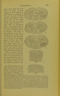
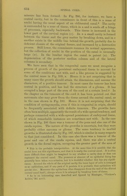
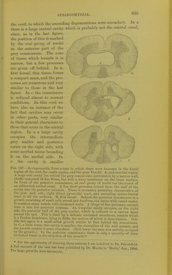
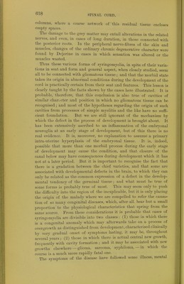
![Sl'INAL CORD. rnissure has been formed. Iu Fig. 186, for instance, we have a centra] cavity, but in the commissure in front of this is a mass of nuclei having the usual aspect of an obliterated canal* The cavity is surrounded by a zone of tissue, which in a and b sends off a fringe ' Processes, especially backwards. This tissue is increased in the lower part of the cervical region (c). In b a small cavity is formed between the tissue and the grey matter by breaking down, and in c another exists in the middle line behind the growth, probably due to deficient closure of the original fissure, and increased by a destructive process. Still lower, the commissure resumes its normal appearance, but the collection of nuclei in the position of the canal is unusually large (d). In the lumbar region the canal is patent (e). The degeneration of the posterior median column and of the lateral columns is secondary. We have seen that in the congenital cases we must recognise a process of growth of the persistent embryonal tissue to account for some of the conditions met with, and a like process is suggested by the central mass in Tig. 109, b. Hence it is not surprising that in many cases the growth should attain the dimensions, and assume the characters, of a positive tumour. In most cases the tumour has been central in position, and has had the structure of a glioma. It has occupied a large part of the area of the cord at a certain level.f In the chapter on the tumours of the cord it has been pointed out that sarcomata also may grow from the tissue around the central canal, as in the case shown in Fig. 181. Hence it is not surprising that the condition of syringomyelia, even if this is congenital in origin, should be frequently associated with definite tumours. The growths have often been multiple, apparently the result of a wide-spread tendency, and perhaps connected with a wide-spi-ead persistence of embryonal tissue, of which remarkable instances are sometimes met with. In the case shown in Pig. 186 there was a tumour of the pons and also one of the cauda equina. The nature of the growths is uncertain, but they were probably either sarcoma or glioma. The same tendency to morbid growths is illustrated also by Fig. 187, which is similar in many respects to that just considered. In this case also there was a tumour of the pons and one of the cauda equina, and there was also a central growth in the dorsal region, occupying the greater part of the area of * This is the probable interpretation. At the same time it is possible that the nuclei have not this significance, and that it is really the central canal. It is often more difficult than might be imagined to say whether a cavity does or does not represent the central canal. The presence or absence of an epithelial lining has generally been taken as a criterion, but it is doubtful whether this has any signifi- cance. Around the whole of the original cavity the inner layer of cells is arranged as an epithelium, and if any part of it persists it is probable that epithelium will persist also. Moreover the epithelium often disappears from the wall of the dilated central canal itself; always where it is enlarged by breaking down of tissue. t As in an interesting case described and figured by Riesinger, ' Virchow's Archiv,' Bd. xcviii.](https://iiif.wellcomecollection.org/image/b21294483_0662.jp2/full/800%2C/0/default.jpg)