A manual of diseases of the nervous system / by Sir W.R. Gowers ; edited by Sir W.R. Gowers and James Taylor.
- William Richard Gowers
- Date:
- 1899
Licence: Public Domain Mark
Credit: A manual of diseases of the nervous system / by Sir W.R. Gowers ; edited by Sir W.R. Gowers and James Taylor. Source: Wellcome Collection.
Provider: This material has been provided by King’s College London. The original may be consulted at King’s College London.
74/720 (page 52)
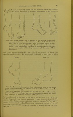
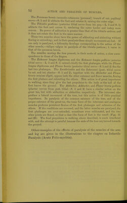
![The Peroneus brevis (musculo.cutaneous [peroneal] branch of ext. popliteal nerve—S. 1 and 2) abducts the Toot and rotates it, raising the outer edge. The Tibial;* -posticus (posterior tibial nerve from int. pop.—L. 5 and S. 1) adducts the Foot and curves it, rendering the outer border and instep more convex, Its power of adduction is greater than that of the tibialis anticus, and it does not rotate the foot in the same manner. These two muscles alone have the power oO adducting and abducting without flexing or extending; and in their paralysis these simple movements are lost, [f one only is paralysed, a deformity develops corresponding to the action of the other muscle,—talipes valgus in paralysis of the tibialis posticus; t, varus in that of the peroneus brevis. The muscles moving the toes present, in their mode of action, a close corre- spondence to those of the fingers. The Extensor loncjus digitorum and the Extensor longus pollicis (anterior tibial nerve —L. 5 and S. 1) extend chiefly the first phalanges, while the Flexor longus digitorum and Flexor brevis (posterior tibial nerve—S. 1 and 2) flex the last two phalanges. The Lumbricales and the Interossei (post, tibial nerve by ext. and int. plantar—S. 1 and 2), together with the Abductor and Flexor brevis minimi digiti, oppose both the other extensor and flexor muscles, flexing the first phalanx and extending the others. This action is of great importance in walking, since they give the last propulsion to the body as the ball of the foot leaves the ground. The Abductor, Adductor, and Flexor brevis pollicis (plantar nerves from post, tibial—S. 1 and 2) have a similar action on the great toe, but with adduction or abduction respectively. The interossei also produce a lateral movement of the toes, but this action is of little practical importance. In paralysis of the common extensor of the toes, and of the proper extensor of the great toe, the tonic force of the interossei and analogous muscles produces persistent flexion of the first phalanges and extension of the others. If the conditions are reversed, and the latter muscles are paralysed, the first phalanges are over-extended, sometimes even subluxated, and the two other joints are flexed, so that a claw-like form of foot is the result (Pigs. 31 and 28). The final propulsion in walking, above described, is much interfered with, and the attempt is painful because the ends of the toes are turned towards the ground. Other examples of the effects of paralysis of the muscles of the arm and leg are given in the illustrations to the chapter on Infantile Paralysis (Acute Polio-myelitis).](https://iiif.wellcomecollection.org/image/b21294483_0078.jp2/full/800%2C/0/default.jpg)