Treatise on Bright's disease of the kidneys : its pathology, diagnosis, and treatment with chapters on the anatomy of the kidney, albuminura and the urinary secretion / by Henry B. Millard.
- Henry Millard
- Date:
- 1892
Licence: Public Domain Mark
Credit: Treatise on Bright's disease of the kidneys : its pathology, diagnosis, and treatment with chapters on the anatomy of the kidney, albuminura and the urinary secretion / by Henry B. Millard. Source: Wellcome Collection.
57/354 (page 31)
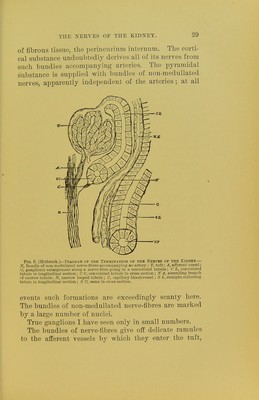
![themselves. Obviously those nerves are most favorable for research which course outside of the epithelia at a small distance from the membrana propria. Here we can, sometimes, see at certain regular intervals, arising at right or acute angles, extremely delicate nerve-fib- rillfe, which pierce the membrana propria and run into the cement substance between the epithelia. The dis- tance in which these ultimate fibrillfe arise fully cor- respond to the breadth of a single epithelial element; so much so that in some places the impression of a lad- der with regular rounds is obtained. Of course, only one of the frames or side-pieces of the ladder is present. In a front view of the epithelia the nerve-fibrillge can sometimes be traced in the form of a delicate plexus dis- tributed in the epithelia, and not infrequent]y convey- ing the impression that every epithelium is surrounded by a nerve-hbrillse in the cement substance. In an edge view this impression is not obtained, for we can see the interstices between the epithelia sup- plied with nerves only exceptionally, while in the ma- jority of cases two or three epithelia seem to be sup- plied with only one nerve-fibrillae common to them. The later image is more particularly pronounced along the straight collecting tubules in which, usually in edge view, two nerve-tibrillse are situated between three or four epithelial elements; and here the cement substance carrjang the nerve-hbrillie is much broader than the cement substance apparently destitute of nerve-fibres. If, however, we recall the fact, that in a front view of the tubules, the arrangement of the ultimate fibrillfe is plexiform, we obviously should not expect to see in edge view nerve-fibrillse between each single epithelium. The distributions of the nerves in the uriniferous tu- bules seem to be richer in the convoluted and the as- cending and descending limbs of the narrow tubules, while the straight collecting ones seem to be more scant-](https://iiif.wellcomecollection.org/image/b20399224_0057.jp2/full/800%2C/0/default.jpg)