Heath's practical anatomy : a manual of dissections / [Christopher Heath].
- Christopher Heath
- Date:
- 1893
Licence: Public Domain Mark
Credit: Heath's practical anatomy : a manual of dissections / [Christopher Heath]. Source: Wellcome Collection.
35/800 page 13
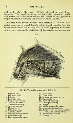
![accompanied by corresponding arterial twigs. Each lateral cuta- neous nerve, except that of the second, divides into anterior and posterior branches, which turn forwards to the pectoral region, and backwards over the latissinius dorsi respectively. The lateral cutaneous branch of the second nerve passes down the inner side of the arm to join the lesser internal cutaneous nerve, or nerve of Wrisberg, and is hence called the intercosto-humeral nerve. The corresponding branch from the third nerve usually accompanies and joins this. These last must be found at once and are to be carefully preserved (Fig. 2, 24). [All the fat is to be removed from the axilla, and the muscles cleaned so far as they are exposed. Care must also be taken not to injure the following structures whilst cleaning them :—the axillary vessels (particularly the vein) and the large nerves, which are to the outer side of the space ; the posterior thoracic nerve, which runs vertically upon the serratus magnus, near the posterior wall of the axilla, giving off a branch to each digitation of the muscle ; the sub- scapular vessels at the outer border of the subscapularis ; the long thoracic artery, near the lower border of the pectoralis minor ; and the three subscapular nerves which cross the space, passing from above downwards and outwards. One or two of the numerous lymphatic glands should also be preserved.] The Axilla (Figs. 2 and 3) is a conical space between the chest and upper arm, and has the following boundaries :—In front, the pectoralis major, the pectoralis minor, and the clavicle and subclavius ; behind, the subscapularis, latissimus dorsi, and teres major muscles, with the body of the scapula; to the i7i7ier side, the serratus magnus, Avith the four upper ribs and intercostal spaces ; and to the outer side, the coracoid process, the upper part of the humerus and the coraco-brachia- lis and biceps muscles. The base of the cone has already been seen to be formed by the axillary fascia stretched between the anterior and posterior boundaries, and the apex is represented by a triangular interval between the clavicle, the upper border of the scapula, and the first rib. The great vessels and nerves, entering at the apex, lie along the outer boundary of the space, the artery being first on the outer side of the nerve cords, then between them, and the vein keeping to the inner side of the artery, whilst most of the branches of the artery take a general direction towards the thorax. Towards the inner side of the space, and imbedded in fat, are the glands, ten or twelve in number, which receive the lymphatics from (1) the front of the chest and the mamma, (2) the abdomen as low as the umbilicus, (3) from the arm and (4) from part of the back. The superficial glands lie along the edge of the pectoralis, and the deeper ones](https://iiif.wellcomecollection.org/image/b20417457_0035.jp2/full/800%2C/0/default.jpg)




 No text description is available for this image
 No text description is available for this image
 No text description is available for this image