Heath's practical anatomy : a manual of dissections / [Christopher Heath].
- Christopher Heath
- Date:
- 1893
Licence: Public Domain Mark
Credit: Heath's practical anatomy : a manual of dissections / [Christopher Heath]. Source: Wellcome Collection.
61/800 page 39
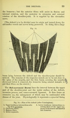
![the humerus ; but the anterior fibres will assist in flexion and internal rotation, and the posterior in extension and external rotation of the shoulder-joint. It is supplied by the circumflex nerve. [The deltoid is to be divided near its origin and turned down, the circumflex vessels and nerve being preserved. In doing this a large Fig. 14. bursa lying between the deltoid and the shoulder-joint should be noticed. The remains of the trapezius muscle are to be cut close to ^ the spine of the scapula, and the thin fascia covering the muscles above and below it removed, the humerus being rotated inwards to put their fibres on the stretch.] The Sub-aeromial Bursd lines the interval between the upper part of the shoulder-joint and the under surface of the deltoid, acromion process, and coraco-acromial ligament. It thus forms an extensive sac, the enlargement of which may be confounded with disease of the joint. Physiologically it represents a synovial lining Fig. 14.—Plan of the deltoid (after Cunningham). 1. Upper tendinous intersections aris- 5. Lower tendinous intersections re- ing from acromion^ ceiving three of the upper seg- 2. Clavicular ffBresT ments of the muscle and inserted 3. Spinous fibres.. into humerus. 4. Lower segments.](https://iiif.wellcomecollection.org/image/b20417457_0061.jp2/full/800%2C/0/default.jpg)


 No text description is available for this image
 No text description is available for this image
 No text description is available for this image